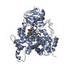
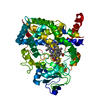
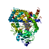
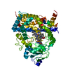
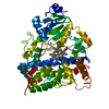
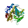
[English] 日本語
Yorodumi
- PDB-6mcw: Crystal structure of the P450 domain of the CYP51-ferredoxin fusi... -

+
Open data


ID or keywords:

Loading...

-
Basic information

Entry
Database: PDB / ID: 6mcw
TitleCrystal structure of the P450 domain of the CYP51-ferredoxin fusion protein from Methylococcus capsulatus, complex with the detergent Anapoe-X-114
ComponentsCytochrome P450 51
KeywordsOXIDOREDUCTASE / CYTOCHROME P450 FOLD / CYP51 / HEME / STEROL BIOSYNTHESIS
Function / homology
Function and homology information


sterol 14alpha-demethylase / steroid 7-alpha-hydroxylase activity / oxysterol 7-alpha-hydroxylase activity / bile acid biosynthetic process / cholesterol homeostasis / iron ion binding / heme binding
Similarity search - Function
4Fe-4S single cluster domain / Cytochrome P450, E-class, group IV / Cytochrome p450 / Cytochrome P450 / 4Fe-4S ferredoxin-type iron-sulfur binding domain profile. / 4Fe-4S ferredoxin-type, iron-sulphur binding domain / Cytochrome P450 / Cytochrome P450 superfamily / Cytochrome P450 / Orthogonal Bundle / Mainly Alpha
Similarity search - Domain/homology
PROTOPORPHYRIN IX CONTAINING FE / Chem-JDJ / Cytochrome P450 51
Similarity search - Component
Biological speciesMethylococcus capsulatus (bacteria)
MethodX-RAY DIFFRACTION / SYNCHROTRON / MOLECULAR REPLACEMENT / Resolution: 2.4 Å
AuthorsHargrove, T. / Wawrzak, Z. / Lamb, D.C. / Lepesheva, G.I.
Funding support United States, 1items
OrganizationGrant numberCountry
National Institutes of Health/National Institute of General Medical Sciences (NIH/NIGMS)R01067871 United States
CitationJournal: Mol.Biol.Evol. / Year: 2020
Title: Concerning P450 evolution: Structural Analyses Support Bacterial Origin of Sterol 14 alpha-Demethylases.
Authors: Lamb, D.C. / Hargrove, T.Y. / Zhao, B. / Wawrzak, Z. / Goldstone, J.V. / Nes, W.D. / Kelly, S.L. / Waterman, M.R. / Stegeman, J.J. / Lepesheva, G.I.
History
DepositionSep 2, 2018Deposition site: RCSB / Processing site: RCSB
Revision 1.0Jul 10, 2019Provider: repository / Type: Initial release
Revision 1.1Jan 1, 2020Group: Author supporting evidence / Category: pdbx_audit_support / Item: _pdbx_audit_support.funding_organization
Revision 1.2Jan 20, 2021Group: Database references / Category: citation / citation_author
Item: _citation.country / _citation.journal_abbrev ..._citation.country / _citation.journal_abbrev / _citation.journal_id_CSD / _citation.journal_id_ISSN / _citation.pdbx_database_id_DOI / _citation.pdbx_database_id_PubMed / _citation.title / _citation.year
Revision 1.3Oct 11, 2023Group: Data collection / Database references / Refinement description
Category: chem_comp_atom / chem_comp_bond ...chem_comp_atom / chem_comp_bond / database_2 / pdbx_initial_refinement_model
Item: _database_2.pdbx_DOI / _database_2.pdbx_database_accession

-
Structure visualization

Structure viewerMolecule:
MolmilJmol/JSmol

Downloads & links

-
Assembly

Deposited unit
A: Cytochrome P450 51
hetero molecules


Theoretical massNumber of molelcules
Total (without water)63,6593
Polymers62,4841
Non-polymers1,1752
Water64936
1


  • Idetical with deposited unit
  • defined by author&software
  • Evidence: gel filtration
TypeNameSymmetry operationNumber
identity operation1_555x,y,z1
MethodPISA
Unit cell
Length a, b, c (Å)150.685, 150.685, 66.321
Angle α, β, γ (deg.)90.00, 90.00, 120.00
Int Tables number152
Space group name H-MP3121

-
Components

#1: Protein Cytochrome P450 51


Mass: 62483.867 Da / Num. of mol.: 1
Source method: isolated from a genetically manipulated source
Source: (gene. exp.) Methylococcus capsulatus (strain ATCC 33009 / NCIMB 11132 / Bath) (bacteria)
Strain: ATCC 33009 / NCIMB 11132 / Bath / Gene: cyp51, MCA2711 / Production host: Escherichia coli (E. coli) / Strain (production host): HMS-164 / References: UniProt: Q603T8, sterol 14alpha-demethylase
#2: Chemical ChemComp-JDJ / 23-[4-(2,4,4-trimethylpentan-2-yl)phenoxy]-3,6,9,12,15,18,21-heptaoxatricosan-1-ol / Anapoe-X-114


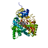
Mass: 558.744 Da / Num. of mol.: 1 / Source method: obtained synthetically / Formula: C30H54O9
#3: Chemical ChemComp-HEM / PROTOPORPHYRIN IX CONTAINING FE / HEME


Mass: 616.487 Da / Num. of mol.: 1 / Source method: obtained synthetically / Formula: C34H32FeN4O4
#4: Water ChemComp-HOH / water


Mass: 18.015 Da / Num. of mol.: 36 / Source method: isolated from a natural source / Formula: H2O

-
Experimental details

-
Experiment

ExperimentMethod: X-RAY DIFFRACTION / Number of used crystals: 1

-
Sample preparation

CrystalDensity Matthews: 3.48 Å3/Da / Density % sol: 64.64 %
Crystal growTemperature: 291 K / Method: vapor diffusion / pH: 7.4 / Details: PEG 6000, photassium phosphate, glycerol, TCEP

-
Data collection

DiffractionMean temperature: 100 K
Diffraction sourceSource: SYNCHROTRON / Site: APS / Beamline: 21-ID-D / Wavelength: 1.07807 Å
DetectorType: MARMOSAIC 300 mm CCD / Detector: CCD / Date: Jun 6, 2018 / Details: FOCUSING MIRRORS
RadiationMonochromator: SI (III) / Protocol: SINGLE WAVELENGTH / Monochromatic (M) / Laue (L): M / Scattering type: x-ray
Radiation wavelengthWavelength: 1.07807 Å / Relative weight: 1
ReflectionResolution: 2.4→30 Å / Num. obs: 33877 / % possible obs: 99 % / Redundancy: 6.2 % / Rmerge(I) obs: 0.054 / Rpim(I) all: 0.043 / Rrim(I) all: 0.109 / Net I/σ(I): 17.6
Reflection shellResolution: 2.4→2.44 Å / Redundancy: 6.4 % / Rmerge(I) obs: 0.873 / Mean I/σ(I) obs: 1.4 / Num. unique obs: 1681 / Rpim(I) all: 0.575 / % possible all: 100

-
Processing

Software
NameVersionClassification
REFMAC5.8.0230refinement
HKL-2000data reduction
HKL-2000data scaling
PHASERphasing
RefinementMethod to determine structure: MOLECULAR REPLACEMENT
Starting model: 6FMO
Resolution: 2.4→29.96 Å / Cor.coef. Fo:Fc: 0.948 / Cor.coef. Fo:Fc free: 0.934 / SU B: 8.609 / SU ML: 0.185 / Cross valid method: THROUGHOUT / ESU R: 0.245 / ESU R Free: 0.197 / Details: HYDROGENS HAVE BEEN ADDED IN THE RIDING POSITIONS
RfactorNum. reflection% reflectionSelection details
Rfree0.23722 1673 4.9 %RANDOM
Rwork0.21582 ---
obs0.21689 32189 99.24 %-
Solvent computationIon probe radii: 0.8 Å / Shrinkage radii: 0.8 Å / VDW probe radii: 1.2 Å
Displacement parametersBiso mean: 58.303 Å2
Baniso -1Baniso -2Baniso -3
1--0.83 Å2-0.41 Å2-0 Å2
2---0.83 Å20 Å2
3---2.68 Å2
Refinement stepCycle: 1 / Resolution: 2.4→29.96 Å
ProteinNucleic acidLigandSolventTotal
Num. atoms3609 0 79 36 3724
Refine LS restraints
Refine-IDTypeDev idealDev ideal targetNumber
X-RAY DIFFRACTIONr_bond_refined_d0.0010.0143781
X-RAY DIFFRACTIONr_bond_other_d00.0173412
X-RAY DIFFRACTIONr_angle_refined_deg0.8211.6965122
X-RAY DIFFRACTIONr_angle_other_deg0.8171.6548006
X-RAY DIFFRACTIONr_dihedral_angle_1_deg8.2565445
X-RAY DIFFRACTIONr_dihedral_angle_2_deg29.99320.045223
X-RAY DIFFRACTIONr_dihedral_angle_3_deg17.60115649
X-RAY DIFFRACTIONr_dihedral_angle_4_deg18.3081542
X-RAY DIFFRACTIONr_chiral_restr0.2530.2469
X-RAY DIFFRACTIONr_gen_planes_refined0.0140.024238
X-RAY DIFFRACTIONr_gen_planes_other0.0060.02702
X-RAY DIFFRACTIONr_nbd_refined
X-RAY DIFFRACTIONr_nbd_other
X-RAY DIFFRACTIONr_nbtor_refined
X-RAY DIFFRACTIONr_nbtor_other
X-RAY DIFFRACTIONr_xyhbond_nbd_refined
X-RAY DIFFRACTIONr_xyhbond_nbd_other
X-RAY DIFFRACTIONr_metal_ion_refined
X-RAY DIFFRACTIONr_metal_ion_other
X-RAY DIFFRACTIONr_symmetry_vdw_refined
X-RAY DIFFRACTIONr_symmetry_vdw_other
X-RAY DIFFRACTIONr_symmetry_hbond_refined
X-RAY DIFFRACTIONr_symmetry_hbond_other
X-RAY DIFFRACTIONr_symmetry_metal_ion_refined
X-RAY DIFFRACTIONr_symmetry_metal_ion_other
X-RAY DIFFRACTIONr_mcbond_it5.3115.6291783
X-RAY DIFFRACTIONr_mcbond_other5.2995.6261782
X-RAY DIFFRACTIONr_mcangle_it7.798.4432227
X-RAY DIFFRACTIONr_mcangle_other7.7888.4472228
X-RAY DIFFRACTIONr_scbond_it6.076.3821996
X-RAY DIFFRACTIONr_scbond_other6.0716.3841994
X-RAY DIFFRACTIONr_scangle_it
X-RAY DIFFRACTIONr_scangle_other8.9619.2832893
X-RAY DIFFRACTIONr_long_range_B_refined12.94467.1064238
X-RAY DIFFRACTIONr_long_range_B_other12.94667.14236
X-RAY DIFFRACTIONr_rigid_bond_restr
X-RAY DIFFRACTIONr_sphericity_free
X-RAY DIFFRACTIONr_sphericity_bonded
LS refinement shellResolution: 2.4→2.462 Å / Total num. of bins used: 20
RfactorNum. reflection% reflection
Rfree0.331 105 -
Rwork0.351 2405 -
obs--99.92 %

+
About Yorodumi

-
News

-
Feb 9, 2022. New format data for meta-information of EMDB entries

New format data for meta-information of EMDB entries

  • Version 3 of the EMDB header file is now the official format.
  • The previous official version 1.9 will be removed from the archive.

Related info.:EMDB header

External links:wwPDB to switch to version 3 of the EMDB data model

-
Aug 12, 2020. Covid-19 info

Covid-19 info

URL: https://pdbjlvh1.pdbj.org/emnavi/covid19.php

New page: Covid-19 featured information page in EM Navigator.

Related info.:Covid-19 info / Mar 5, 2020. Novel coronavirus structure data

+
Mar 5, 2020. Novel coronavirus structure data

Novel coronavirus structure data

Related info.:Yorodumi Speices / Aug 12, 2020. Covid-19 info

External links:COVID-19 featured content - PDBj / Molecule of the Month (242):Coronavirus Proteases

+
Jan 31, 2019. EMDB accession codes are about to change! (news from PDBe EMDB page)

EMDB accession codes are about to change! (news from PDBe EMDB page)

  • The allocation of 4 digits for EMDB accession codes will soon come to an end. Whilst these codes will remain in use, new EMDB accession codes will include an additional digit and will expand incrementally as the available range of codes is exhausted. The current 4-digit format prefixed with “EMD-” (i.e. EMD-XXXX) will advance to a 5-digit format (i.e. EMD-XXXXX), and so on. It is currently estimated that the 4-digit codes will be depleted around Spring 2019, at which point the 5-digit format will come into force.
  • The EM Navigator/Yorodumi systems omit the EMD- prefix.

Related info.:Q: What is EMD? / ID/Accession-code notation in Yorodumi/EM Navigator

External links:EMDB Accession Codes are Changing Soon! / Contact to PDBj

+
Jul 12, 2017. Major update of PDB

Major update of PDB

  • wwPDB released updated PDB data conforming to the new PDBx/mmCIF dictionary.
  • This is a major update changing the version number from 4 to 5, and with Remediation, in which all the entries are updated.
  • In this update, many items about electron microscopy experimental information are reorganized (e.g. em_software).
  • Now, EM Navigator and Yorodumi are based on the updated data.

External links:wwPDB Remediation / Enriched Model Files Conforming to OneDep Data Standards Now Available in the PDB FTP Archive

-
Yorodumi

Thousand views of thousand structures

  • Yorodumi is a browser for structure data from EMDB, PDB, SASBDB, etc.
  • This page is also the successor to EM Navigator detail page, and also detail information page/front-end page for Omokage search.
  • The word "yorodu" (or yorozu) is an old Japanese word meaning "ten thousand". "mi" (miru) is to see.

Related info.:EMDB / PDB / SASBDB / Comparison of 3 databanks / Yorodumi Search / Aug 31, 2016. New EM Navigator & Yorodumi / Yorodumi Papers / Jmol/JSmol / Function and homology information / Changes in new EM Navigator and Yorodumi

Read more