 Movie
Movie Controller

[English] 日本語
 Yorodumi
- PDB-6m6p: Structure of Marine bacterial laminarinase mutant E135A in comple... -
+ Open data

- Basic information
| Entry | Database: PDB / ID: 6m6p | ||||||
|---|---|---|---|---|---|---|---|
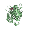
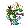
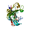
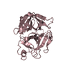
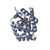
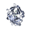
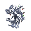
| Title | Structure of Marine bacterial laminarinase mutant E135A in complex with 1,3-beta-cellotriosyl-glucose | ||||||
 Components Components | laminarinase | ||||||
 Keywords Keywords | HYDROLASE | ||||||
| Function / homology |  Function and homology information Function and homology informationhydrolase activity, hydrolyzing O-glycosyl compounds / carbohydrate binding / carbohydrate metabolic process / metal ion binding Similarity search - Function | ||||||
| Biological species |  Aquimarina sp. (bacteria) Aquimarina sp. (bacteria) | ||||||
| Method |  X-RAY DIFFRACTION / SYNCHROTRON / MOLECULAR REPLACEMENT / Resolution: 2.27 Å X-RAY DIFFRACTION / SYNCHROTRON / MOLECULAR REPLACEMENT / Resolution: 2.27 Å | ||||||
 Authors Authors | Yang, J. / Xu, Y. / Tanokura, M. / Long, L. / Miyakawa, T. | ||||||
 Citation Citation | Journal: Appl.Environ.Microbiol. / Year: 2020 Title: Molecular Basis for Substrate Recognition and Catalysis by a Marine Bacterial Laminarinase. Authors: Yang, J. / Xu, Y. / Miyakawa, T. / Long, L. / Tanokura, M. | ||||||
| History |
|
- Structure visualization
| Structure viewer | Molecule: MolmilJmol/JSmol |
|---|
- Downloads & links
-Download
| PDBx/mmCIF format | 6m6p.cif.gz | 67.4 KB | Display | PDBx/mmCIF format |
|---|---|---|---|---|
| PDB format | pdb6m6p.ent.gz | 46.6 KB | Display | PDB format |
| PDBx/mmJSON format | 6m6p.json.gz | Tree view | PDBx/mmJSON format | |
| Others |  Other downloads Other downloads |
-Validation report
| Arichive directory | https://data.pdbj.org/pub/pdb/validation_reports/m6/6m6pftp://data.pdbj.org/pub/pdb/validation_reports/m6/6m6p | HTTPS FTP |
|---|
-Related structure data
| Related structure data |  6jh5C  6jhjC  6jiaC  4crqS C: citing same article ( S: Starting model for refinement |
|---|---|
| Similar structure data |


-Links
 PDBj
PDBj

- Assembly
| Deposited unit | 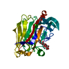
| ||||||||
|---|---|---|---|---|---|---|---|---|---|
| 1 |
| ||||||||
| Unit cell |
| ||||||||
| Components on special symmetry positions |
|
-Components
| #1: Protein | Mass: 27776.602 Da / Num. of mol.: 1 / Mutation: E135A Source method: isolated from a genetically manipulated source Source: (gene. exp.) Aquimarina sp. (bacteria) / Production host: |
|---|---|
| #2: Polysaccharide | beta-D-glucopyranose-(1-4)-beta-D-glucopyranose-(1-4)-beta-D-glucopyranose-(1-3)-alpha-D-glucopyranose Source method: isolated from a genetically manipulated source |
| #3: Chemical | ChemComp-CA /   Mass: 40.078 Da / Num. of mol.: 1 / Source method: obtained synthetically / Formula: Ca / Feature type: SUBJECT OF INVESTIGATION Mass: 40.078 Da / Num. of mol.: 1 / Source method: obtained synthetically / Formula: Ca / Feature type: SUBJECT OF INVESTIGATION |
| #4: Water | ChemComp-HOH /  Mass: 18.015 Da / Num. of mol.: 121 / Source method: isolated from a natural source / Formula: H2O Mass: 18.015 Da / Num. of mol.: 121 / Source method: isolated from a natural source / Formula: H2O |
| Has ligand of interest | Y |
| Sequence details | This protein is E135A mutant. |
-Experimental details
-Experiment
| Experiment | Method: X-RAY DIFFRACTION / Number of used crystals: 1 |
|---|
- Sample preparation
| Crystal | Density Matthews: 2.1 Å3/Da / Density % sol: 41.44 % |
|---|---|
| Crystal grow | Temperature: 293 K / Method: vapor diffusion, sitting drop Details: 100 mM MES (pH7.0), 15% (v/v) ethanol, 28% (m/v) PEG20000 |
-Data collection
| Diffraction | Mean temperature: 95 K / Serial crystal experiment: N | ||||||||||||||||||||||||
|---|---|---|---|---|---|---|---|---|---|---|---|---|---|---|---|---|---|---|---|---|---|---|---|---|---|
| Diffraction source | Source: SYNCHROTRON / Site: Photon Factory  / Beamline: BL-5A / Wavelength: 1 Å / Beamline: BL-5A / Wavelength: 1 Å | ||||||||||||||||||||||||
| Detector | Type: DECTRIS PILATUS3 S 2M / Detector: PIXEL / Date: Nov 17, 2018 | ||||||||||||||||||||||||
| Radiation | Protocol: SINGLE WAVELENGTH / Monochromatic (M) / Laue (L): M / Scattering type: x-ray | ||||||||||||||||||||||||
| Radiation wavelength | Wavelength: 1 Å / Relative weight: 1 | ||||||||||||||||||||||||
| Reflection | Resolution: 2.21→47.82 Å / Num. obs: 11550 / % possible obs: 95.9 % / Redundancy: 6.4 % / CC1/2: 0.997 / Rmerge(I) obs: 0.107 / Rpim(I) all: 0.046 / Rrim(I) all: 0.117 / Net I/σ(I): 10.4 / Num. measured all: 74218 / Scaling rejects: 9 | ||||||||||||||||||||||||
| Reflection shell | Diffraction-ID: 1
|
- Processing
| Software |
| ||||||||||||||||||||||||||||||||||||||||||||||||||||||||||||
|---|---|---|---|---|---|---|---|---|---|---|---|---|---|---|---|---|---|---|---|---|---|---|---|---|---|---|---|---|---|---|---|---|---|---|---|---|---|---|---|---|---|---|---|---|---|---|---|---|---|---|---|---|---|---|---|---|---|---|---|---|---|
| Refinement | Method to determine structure: MOLECULAR REPLACEMENT Starting model: 4CRQ Resolution: 2.27→43.9 Å / Cor.coef. Fo:Fc: 0.96 / Cor.coef. Fo:Fc free: 0.927 / SU B: 6.981 / SU ML: 0.164 / Cross valid method: THROUGHOUT / σ(F): 0 / ESU R: 0.38 / ESU R Free: 0.234 / Stereochemistry target values: MAXIMUM LIKELIHOOD Details: HYDROGENS HAVE BEEN ADDED IN THE RIDING POSITIONS U VALUES : REFINED INDIVIDUALLY
| ||||||||||||||||||||||||||||||||||||||||||||||||||||||||||||
| Solvent computation | Ion probe radii: 0.8 Å / Shrinkage radii: 0.8 Å / VDW probe radii: 1.2 Å / Solvent model: MASK | ||||||||||||||||||||||||||||||||||||||||||||||||||||||||||||
| Displacement parameters | Biso max: 105.99 Å2 / Biso mean: 33.324 Å2 / Biso min: 21.43 Å2
| ||||||||||||||||||||||||||||||||||||||||||||||||||||||||||||
| Refinement step | Cycle: final / Resolution: 2.27→43.9 Å
| ||||||||||||||||||||||||||||||||||||||||||||||||||||||||||||
| Refine LS restraints |
| ||||||||||||||||||||||||||||||||||||||||||||||||||||||||||||
| LS refinement shell | Resolution: 2.271→2.33 Å / Rfactor Rfree error: 0 / Total num. of bins used: 20
|
