 Movie
Movie Controller

[English] 日本語
 Yorodumi
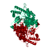
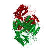
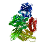
- PDB-6m5a: Crystal structure of GH121 beta-L-arabinobiosidase HypBA2 from Bi... -
+ Open data

- Basic information
| Entry | Database: PDB / ID: 6m5a | |||||||||||||||
|---|---|---|---|---|---|---|---|---|---|---|---|---|---|---|---|---|
| Title | Crystal structure of GH121 beta-L-arabinobiosidase HypBA2 from Bifidobacterium longum | |||||||||||||||
 Components Components | Beta-L-arabinobiosidase | |||||||||||||||
 Keywords Keywords | HYDROLASE / (alpha/alpha)6 barrel / glycoside hydrolase family 121 | |||||||||||||||
| Function / homology |  Function and homology information Function and homology information(Ara-f)3-Hyp beta-L-arabinobiosidase / hydrolase activity, acting on glycosyl bonds / polysaccharide catabolic process / membrane => GO:0016020 / carbohydrate metabolic process / membrane Similarity search - Function | |||||||||||||||
| Biological species |  Bifidobacterium longum (bacteria) Bifidobacterium longum (bacteria) | |||||||||||||||
| Method |  X-RAY DIFFRACTION / SYNCHROTRON / SAD / Resolution: 1.85 Å X-RAY DIFFRACTION / SYNCHROTRON / SAD / Resolution: 1.85 Å | |||||||||||||||
 Authors Authors | Saito, K. / Arakawa, T. / Yamada, C. / Fujita, K. / Fushinobu, S. | |||||||||||||||
| Funding support |  Japan, 4items Japan, 4items
| |||||||||||||||
 Citation Citation | Journal: Plos One / Year: 2020 Title: Crystal structure of beta-L-arabinobiosidase belonging to glycoside hydrolase family 121. Authors: Saito, K. / Viborg, A.H. / Sakamoto, S. / Arakawa, T. / Yamada, C. / Fujita, K. / Fushinobu, S. #1: Journal: J. Biol. Chem. / Year: 2011Title: Molecular cloning and characterization of a beta-L-Arabinobiosidase in Bifidobacterium longum that belongs to a novel glycoside hydrolase family. Authors: Fujita, K. / Sakamoto, S. / Ono, Y. / Wakao, M. / Suda, Y. / Kitahara, K. / Suganuma, T. | |||||||||||||||
| History |
|
- Structure visualization
| Structure viewer | Molecule: MolmilJmol/JSmol |
|---|
- Downloads & links
-Download
| PDBx/mmCIF format | 6m5a.cif.gz | 200.5 KB | Display | PDBx/mmCIF format |
|---|---|---|---|---|
| PDB format | pdb6m5a.ent.gz | 151.7 KB | Display | PDB format |
| PDBx/mmJSON format | 6m5a.json.gz | Tree view | PDBx/mmJSON format | |
| Others |  Other downloads Other downloads |
-Validation report
| Arichive directory | https://data.pdbj.org/pub/pdb/validation_reports/m5/6m5aftp://data.pdbj.org/pub/pdb/validation_reports/m5/6m5a | HTTPS FTP |
|---|
-Related structure data
| Similar structure data |
|---|

-Links
 PDBj
PDBj


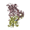
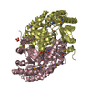
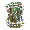
- Assembly
| Deposited unit | 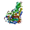
| ||||||||
|---|---|---|---|---|---|---|---|---|---|
| 1 |
| ||||||||
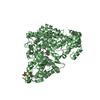
| Unit cell |
|
-Components
-Protein , 1 types, 1 molecules A
| #1: Protein | Mass: 96944.609 Da / Num. of mol.: 1 / Fragment: catalytic domain Source method: isolated from a genetically manipulated source Source: (gene. exp.) Bifidobacterium longum (bacteria) / Gene: DWV93_04865 / Plasmid: pET-23d / Production host: |
|---|
-Non-polymers , 6 types, 575 molecules 


| #2: Chemical | ChemComp-PEG /  Mass: 106.120 Da / Num. of mol.: 8 / Source method: obtained synthetically / Formula: C4H10O3 Mass: 106.120 Da / Num. of mol.: 8 / Source method: obtained synthetically / Formula: C4H10O3#3: Chemical | ChemComp-PGE /  Mass: 150.173 Da / Num. of mol.: 4 / Source method: obtained synthetically / Formula: C6H14O4 Mass: 150.173 Da / Num. of mol.: 4 / Source method: obtained synthetically / Formula: C6H14O4#4: Chemical | ChemComp-EDO /  Mass: 62.068 Da / Num. of mol.: 10 / Source method: obtained synthetically / Formula: C2H6O2 Mass: 62.068 Da / Num. of mol.: 10 / Source method: obtained synthetically / Formula: C2H6O2#5: Chemical | ChemComp-PG4 / |  Mass: 194.226 Da / Num. of mol.: 1 / Source method: obtained synthetically / Formula: C8H18O5 / Comment: precipitant*YM Mass: 194.226 Da / Num. of mol.: 1 / Source method: obtained synthetically / Formula: C8H18O5 / Comment: precipitant*YM#6: Chemical |  Mass: 40.078 Da / Num. of mol.: 3 / Source method: obtained synthetically / Formula: Ca Mass: 40.078 Da / Num. of mol.: 3 / Source method: obtained synthetically / Formula: Ca#7: Water | ChemComp-HOH / | Mass: 18.015 Da / Num. of mol.: 549 / Source method: isolated from a natural source / Formula: H2O |
|---|
-Details
| Has ligand of interest | N |
|---|
-Experimental details
-Experiment
| Experiment | Method: X-RAY DIFFRACTION / Number of used crystals: 1 |
|---|
- Sample preparation
| Crystal | Density Matthews: 2.39 Å3/Da / Density % sol: 48.43 % / Description: Pillar |
|---|---|
| Crystal grow | Temperature: 293 K / Method: vapor diffusion, sitting drop / pH: 6.6 / Details: 17% (w/v) PEG1000 and 0.1 M Tris-HCl (pH 6.6) |
-Data collection
| Diffraction | Mean temperature: 100 K / Serial crystal experiment: N |
|---|---|
| Diffraction source | Source: SYNCHROTRON / Site: Photon Factory / Beamline: BL-1A / Wavelength: 1.1 Å |
| Detector | Type: DECTRIS EIGER X 4M / Detector: PIXEL / Date: Nov 4, 2016 |
| Radiation | Monochromator: Cryo-cooled channel-cut Si(111) / Protocol: SINGLE WAVELENGTH / Monochromatic (M) / Laue (L): M / Scattering type: x-ray |
| Radiation wavelength | Wavelength: 1.1 Å / Relative weight: 1 |
| Reflection | Resolution: 1.85→44.35 Å / Num. obs: 79741 / % possible obs: 100 % / Redundancy: 6.8 % / Biso Wilson estimate: 21.018 Å2 / CC1/2: 0.94 / Rmerge(I) obs: 0.123 / Rpim(I) all: 0.051 / Χ2: 0.86 / Net I/σ(I): 9.1 |
| Reflection shell | Resolution: 1.85→1.89 Å / Redundancy: 7 % / Rmerge(I) obs: 1.405 / Mean I/σ(I) obs: 1.3 / Num. unique obs: 4473 / CC1/2: 0.634 / Rpim(I) all: 0.571 / Χ2: 0.83 / % possible all: 100 |
- Processing
| Software |
| ||||||||||||||||||||||||||||||||||||||||||||||||||||||||||||
|---|---|---|---|---|---|---|---|---|---|---|---|---|---|---|---|---|---|---|---|---|---|---|---|---|---|---|---|---|---|---|---|---|---|---|---|---|---|---|---|---|---|---|---|---|---|---|---|---|---|---|---|---|---|---|---|---|---|---|---|---|---|
| Refinement | Method to determine structure: SAD / Resolution: 1.85→44.35 Å / Cor.coef. Fo:Fc: 0.966 / Cor.coef. Fo:Fc free: 0.957 / SU B: 3.065 / SU ML: 0.087 / Cross valid method: THROUGHOUT / σ(F): 0 / ESU R: 0.118 / ESU R Free: 0.109 Details: HYDROGENS HAVE BEEN ADDED IN THE RIDING POSITIONS U VALUES : REFINED INDIVIDUALLY
| ||||||||||||||||||||||||||||||||||||||||||||||||||||||||||||
| Solvent computation | Ion probe radii: 0.8 Å / Shrinkage radii: 0.8 Å / VDW probe radii: 1.2 Å | ||||||||||||||||||||||||||||||||||||||||||||||||||||||||||||
| Displacement parameters | Biso max: 87.76 Å2 / Biso mean: 27.5 Å2 / Biso min: 17.32 Å2
| ||||||||||||||||||||||||||||||||||||||||||||||||||||||||||||
| Refinement step | Cycle: final / Resolution: 1.85→44.35 Å
| ||||||||||||||||||||||||||||||||||||||||||||||||||||||||||||
| Refine LS restraints |
| ||||||||||||||||||||||||||||||||||||||||||||||||||||||||||||
| LS refinement shell | Resolution: 1.85→1.898 Å / Rfactor Rfree error: 0 / Total num. of bins used: 20
|
