 Movie
Movie Controller


+ Open data

- Basic information
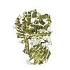
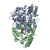
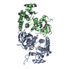
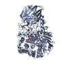
| Entry | Database: PDB / ID: 6m4j | ||||||
|---|---|---|---|---|---|---|---|
| Title | SspA in complex with cysteine | ||||||
 Components Components | SspA complex protein | ||||||
 Keywords Keywords | OXIDOREDUCTASE / cysteine desulfhydrase | ||||||
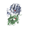
| Function / homology | Aspartate Aminotransferase; domain 2 / Type I PLP-dependent aspartate aminotransferase-like (Major domain) / 3-Layer(aba) Sandwich / Alpha Beta / CYSTEINE / PYRIDOXAL-5'-PHOSPHATE Function and homology information Function and homology information | ||||||
| Biological species |  Vibrio cyclitrophicus (bacteria) Vibrio cyclitrophicus (bacteria) | ||||||
| Method |  X-RAY DIFFRACTION / SYNCHROTRON / MOLECULAR REPLACEMENT / Resolution: 1.8 Å X-RAY DIFFRACTION / SYNCHROTRON / MOLECULAR REPLACEMENT / Resolution: 1.8 Å | ||||||
 Authors Authors | Liu, L. / Gao, H. | ||||||
 Citation Citation | Journal: Mbio / Year: 2020 Title: Structural Analysis of an l-Cysteine Desulfurase from an Ssp DNA Phosphorothioation System. Authors: Liu, L. / Jiang, S. / Xing, M. / Chen, C. / Lai, C. / Li, N. / Liu, G. / Wu, D. / Gao, H. / Hong, L. / Tan, P. / Chen, S. / Deng, Z. / Wu, G. / Wang, L. | ||||||
| History |
|
- Structure visualization
| Structure viewer | Molecule: MolmilJmol/JSmol |
|---|
- Downloads & links
-Download
| PDBx/mmCIF format | 6m4j.cif.gz | 313.4 KB | Display | PDBx/mmCIF format |
|---|---|---|---|---|
| PDB format | pdb6m4j.ent.gz | 254.8 KB | Display | PDB format |
| PDBx/mmJSON format | 6m4j.json.gz | Tree view | PDBx/mmJSON format | |
| Others |  Other downloads Other downloads |
-Validation report
| Arichive directory | https://data.pdbj.org/pub/pdb/validation_reports/m4/6m4jftp://data.pdbj.org/pub/pdb/validation_reports/m4/6m4j | HTTPS FTP |
|---|
-Related structure data
| Related structure data | 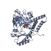 3vaxS S: Starting model for refinement |
|---|---|
| Similar structure data |


-Links
 PDBj
PDBj

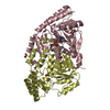
- Assembly
| Deposited unit | 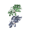
| ||||||||||||
|---|---|---|---|---|---|---|---|---|---|---|---|---|---|
| 1 | 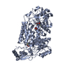
| ||||||||||||
| 2 | 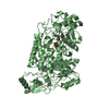
| ||||||||||||
| Unit cell |
| ||||||||||||
| Components on special symmetry positions |
| ||||||||||||
| Noncrystallographic symmetry (NCS) | NCS oper:
|
-Components
| #1: Protein | Mass: 37897.465 Da / Num. of mol.: 2 / Mutation: C314S Source method: isolated from a genetically manipulated source Source: (gene. exp.) Vibrio cyclitrophicus (bacteria) / Production host: #2: Chemical |   Mass: 247.142 Da / Num. of mol.: 2 / Source method: obtained synthetically / Formula: C8H10NO6P / Feature type: SUBJECT OF INVESTIGATION Mass: 247.142 Da / Num. of mol.: 2 / Source method: obtained synthetically / Formula: C8H10NO6P / Feature type: SUBJECT OF INVESTIGATION#3: Chemical |   Type: L-peptide linking / Mass: 121.158 Da / Num. of mol.: 2 / Source method: obtained synthetically / Formula: C3H7NO2S / Feature type: SUBJECT OF INVESTIGATION Type: L-peptide linking / Mass: 121.158 Da / Num. of mol.: 2 / Source method: obtained synthetically / Formula: C3H7NO2S / Feature type: SUBJECT OF INVESTIGATION#4: Water | ChemComp-HOH / |  Mass: 18.015 Da / Num. of mol.: 863 / Source method: isolated from a natural source / Formula: H2O Mass: 18.015 Da / Num. of mol.: 863 / Source method: isolated from a natural source / Formula: H2OHas ligand of interest | Y | Sequence details | The sequence has been deposited to NCBI with accession code WP_016789103.1. And C314S mutation was introduced. | |
|---|
-Experimental details
-Experiment
| Experiment | Method: X-RAY DIFFRACTION / Number of used crystals: 1 |
|---|
- Sample preparation
| Crystal | Density Matthews: 2.46 Å3/Da / Density % sol: 50.05 % |
|---|---|
| Crystal grow | Temperature: 287.15 K / Method: vapor diffusion, hanging drop / pH: 7 / Details: 1.8M ammonium citrate, pH 7.0 / PH range: 6.5-7.5 |
-Data collection
| Diffraction | Mean temperature: 173 K / Serial crystal experiment: N |
|---|---|
| Diffraction source | Source: SYNCHROTRON / Site: SSRF  / Beamline: BL17U / Wavelength: 0.9789 Å / Beamline: BL17U / Wavelength: 0.9789 Å |
| Detector | Type: DECTRIS PILATUS 2M / Detector: PIXEL / Date: Apr 1, 2016 |
| Radiation | Protocol: MAD / Monochromatic (M) / Laue (L): M / Scattering type: x-ray |
| Radiation wavelength | Wavelength: 0.9789 Å / Relative weight: 1 |
| Reflection | Resolution: 1.8→115 Å / Num. obs: 72776 / % possible obs: 100 % / Redundancy: 12.5 % / CC1/2: 0.962 / Net I/σ(I): 5.3 |
| Reflection shell | Resolution: 1.8→1.84 Å / Num. unique obs: 11922 / CC1/2: 0.962 |
- Processing
| Software |
| ||||||||||||||||||||||||||||||||||||||||||||||||||||||||||||
|---|---|---|---|---|---|---|---|---|---|---|---|---|---|---|---|---|---|---|---|---|---|---|---|---|---|---|---|---|---|---|---|---|---|---|---|---|---|---|---|---|---|---|---|---|---|---|---|---|---|---|---|---|---|---|---|---|---|---|---|---|---|
| Refinement | Method to determine structure: MOLECULAR REPLACEMENT Starting model: 3VAX Resolution: 1.8→114.54 Å / Cor.coef. Fo:Fc: 0.963 / Cor.coef. Fo:Fc free: 0.953 / SU B: 5.106 / SU ML: 0.071 / Cross valid method: THROUGHOUT / σ(F): 0 / ESU R: 0.219 / ESU R Free: 0.106 Details: HYDROGENS HAVE BEEN USED IF PRESENT IN THE INPUT U VALUES : REFINED INDIVIDUALLY
| ||||||||||||||||||||||||||||||||||||||||||||||||||||||||||||
| Solvent computation | Ion probe radii: 0.8 Å / Shrinkage radii: 0.8 Å / VDW probe radii: 1.2 Å | ||||||||||||||||||||||||||||||||||||||||||||||||||||||||||||
| Displacement parameters | Biso max: 63.79 Å2 / Biso mean: 22.156 Å2 / Biso min: 12.27 Å2
| ||||||||||||||||||||||||||||||||||||||||||||||||||||||||||||
| Refinement step | Cycle: final / Resolution: 1.8→114.54 Å
| ||||||||||||||||||||||||||||||||||||||||||||||||||||||||||||
| Refine LS restraints |
| ||||||||||||||||||||||||||||||||||||||||||||||||||||||||||||
| Refine LS restraints NCS | Number: 537 / Type: TIGHT THERMAL / Rms dev position: 3.24 Å / Weight position: 0.5 | ||||||||||||||||||||||||||||||||||||||||||||||||||||||||||||
| LS refinement shell | Resolution: 1.801→1.848 Å / Rfactor Rfree error: 0 / Total num. of bins used: 20
|
