 Movie
Movie Controller

[English] 日本語
 Yorodumi
- PDB-6m37: The crystal structure of B. subtilis RsbV/RsbW complex in the hex... -
+ Open data

- Basic information
| Entry | Database: PDB / ID: 6m37 | |||||||||
|---|---|---|---|---|---|---|---|---|---|---|
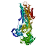
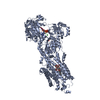
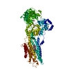
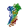
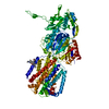
| Title | The crystal structure of B. subtilis RsbV/RsbW complex in the hexagonal crystal form | |||||||||
 Components Components |
| |||||||||
 Keywords Keywords | SIGNALING PROTEIN / sigma factor / anti-sigma / anti-anti-sigma / SigB / RsbW / RsbV | |||||||||
| Function / homology |  Function and homology information Function and homology informationanti-sigma factor antagonist activity / sigma factor antagonist activity / non-specific serine/threonine protein kinase / protein serine kinase activity / negative regulation of DNA-templated transcription / protein serine/threonine kinase activity / ATP binding Similarity search - Function | |||||||||
| Biological species |  | |||||||||
| Method |  X-RAY DIFFRACTION / SYNCHROTRON / MOLECULAR REPLACEMENT / Resolution: 3.1 Å X-RAY DIFFRACTION / SYNCHROTRON / MOLECULAR REPLACEMENT / Resolution: 3.1 Å | |||||||||
 Authors Authors | Pathak, D. / Kwon, E. / Kim, D.Y. | |||||||||
| Funding support |  Korea, Republic Of, 2items Korea, Republic Of, 2items
| |||||||||
 Citation Citation | Journal: Iucrj / Year: 2020 Title: Structural insights into the regulation of SigB activity by RsbV and RsbW. Authors: Pathak, D. / Jin, K.S. / Tandukar, S. / Kim, J.H. / Kwon, E. / Kim, D.Y. | |||||||||
| History |
|
- Structure visualization
| Structure viewer | Molecule: MolmilJmol/JSmol |
|---|
- Downloads & links
-Download
| PDBx/mmCIF format | 6m37.cif.gz | 95 KB | Display | PDBx/mmCIF format |
|---|---|---|---|---|
| PDB format | pdb6m37.ent.gz | 72.6 KB | Display | PDB format |
| PDBx/mmJSON format | 6m37.json.gz | Tree view | PDBx/mmJSON format | |
| Others |  Other downloads Other downloads |
-Validation report
| Arichive directory | https://data.pdbj.org/pub/pdb/validation_reports/m3/6m37ftp://data.pdbj.org/pub/pdb/validation_reports/m3/6m37 | HTTPS FTP |
|---|
-Related structure data
| Related structure data |  6m36C  1thnS 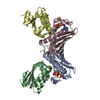 1tilS S: Starting model for refinement C: citing same article ( |
|---|---|
| Similar structure data |

-Links
 PDBj
PDBj
- Assembly
| Deposited unit | 
| ||||||||||||||||||||||||||||||||||||||||||||||||||||||||||||||||||||||||||||||||||||||||||
|---|---|---|---|---|---|---|---|---|---|---|---|---|---|---|---|---|---|---|---|---|---|---|---|---|---|---|---|---|---|---|---|---|---|---|---|---|---|---|---|---|---|---|---|---|---|---|---|---|---|---|---|---|---|---|---|---|---|---|---|---|---|---|---|---|---|---|---|---|---|---|---|---|---|---|---|---|---|---|---|---|---|---|---|---|---|---|---|---|---|---|---|
| 1 | 
| ||||||||||||||||||||||||||||||||||||||||||||||||||||||||||||||||||||||||||||||||||||||||||
| Unit cell |
| ||||||||||||||||||||||||||||||||||||||||||||||||||||||||||||||||||||||||||||||||||||||||||
| Noncrystallographic symmetry (NCS) | NCS domain:
NCS domain segments:
NCS ensembles :
|
-Components
| #1: Protein | Mass: 15702.462 Da / Num. of mol.: 2 Source method: isolated from a genetically manipulated source Source: (gene. exp.) Strain: 168 / Gene: rsbW, BSU04720 / Production host: References: UniProt: P17904, non-specific serine/threonine protein kinase #2: Protein | Mass: 11350.083 Da / Num. of mol.: 2 Source method: isolated from a genetically manipulated source Source: (gene. exp.) Strain: 168 / Gene: rsbV, BSU04710 / Production host: |
|---|
-Experimental details
-Experiment
| Experiment | Method: X-RAY DIFFRACTION / Number of used crystals: 1 |
|---|
- Sample preparation
| Crystal | Density Matthews: 3.22 Å3/Da / Density % sol: 61.86 % / Description: Rod-shaped hexagonal crystal |
|---|---|
| Crystal grow | Temperature: 293 K / Method: batch mode / Details: 20% w/v PEG3350 and 200 mM Potassium formate, ADP |
-Data collection
| Diffraction | Mean temperature: 80 K / Serial crystal experiment: N |
|---|---|
| Diffraction source | Source: SYNCHROTRON / Site: PAL/PLS / Beamline: 11C / Wavelength: 0.97933 Å |
| Detector | Type: DECTRIS PILATUS3 6M / Detector: PIXEL / Date: Sep 2, 2019 |
| Radiation | Protocol: SINGLE WAVELENGTH / Monochromatic (M) / Laue (L): M / Scattering type: x-ray |
| Radiation wavelength | Wavelength: 0.97933 Å / Relative weight: 1 |
| Reflection | Resolution: 3.1→50 Å / Num. obs: 13426 / % possible obs: 99.9 % / Redundancy: 9.5 % / Biso Wilson estimate: 87.6 Å2 / Rmerge(I) obs: 0.09 / Net I/σ(I): 15.3 |
| Reflection shell | Resolution: 3.1→3.31 Å / Rmerge(I) obs: 0.654 / Mean I/σ(I) obs: 3.6 / Num. unique obs: 2377 |
- Processing
| Software |
| |||||||||||||||||||||||||||||||||||
|---|---|---|---|---|---|---|---|---|---|---|---|---|---|---|---|---|---|---|---|---|---|---|---|---|---|---|---|---|---|---|---|---|---|---|---|---|
| Refinement | Method to determine structure: MOLECULAR REPLACEMENT Starting model: 1THN, 1TIL Resolution: 3.1→45.668 Å / SU ML: 0.37 / Cross valid method: THROUGHOUT / σ(F): 1.34 / Phase error: 26.85 / Stereochemistry target values: ML
| |||||||||||||||||||||||||||||||||||
| Solvent computation | Shrinkage radii: 0.9 Å / VDW probe radii: 1.11 Å / Solvent model: FLAT BULK SOLVENT MODEL | |||||||||||||||||||||||||||||||||||
| Displacement parameters | Biso max: 155.25 Å2 / Biso mean: 80.5029 Å2 / Biso min: 41.93 Å2 | |||||||||||||||||||||||||||||||||||
| Refinement step | Cycle: final / Resolution: 3.1→45.668 Å
| |||||||||||||||||||||||||||||||||||
| Refine LS restraints NCS |
| |||||||||||||||||||||||||||||||||||
| LS refinement shell | Refine-ID: X-RAY DIFFRACTION / Rfactor Rfree error: 0 / % reflection obs: 100 %
|
