 Movie
Movie Controller

[English] 日本語
 Yorodumi
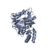
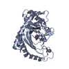
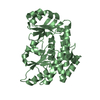
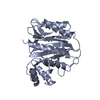
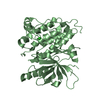
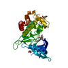
- PDB-6m00: crystal structure of Methionine aminopeptidase from Pyrococcus fu... -
+ Open data

- Basic information
| Entry | Database: PDB / ID: 6m00 | ||||||
|---|---|---|---|---|---|---|---|
| Title | crystal structure of Methionine aminopeptidase from Pyrococcus furiosus | ||||||
 Components Components | Methionine aminopeptidase | ||||||
 Keywords Keywords | HYDROLASE / Methionine aminopeptidase | ||||||
| Function / homology |  Function and homology information Function and homology informationinitiator methionyl aminopeptidase activity / methionyl aminopeptidase / metalloaminopeptidase activity / proteolysis / metal ion binding / cytoplasm Similarity search - Function | ||||||
| Biological species |   Pyrococcus furiosus (archaea) Pyrococcus furiosus (archaea) | ||||||
| Method |  X-RAY DIFFRACTION / SYNCHROTRON / MOLECULAR REPLACEMENT / molecular replacement / Resolution: 3.2 Å X-RAY DIFFRACTION / SYNCHROTRON / MOLECULAR REPLACEMENT / molecular replacement / Resolution: 3.2 Å | ||||||
 Authors Authors | Sandeep, C.B. / Addlagatta, A. | ||||||
| Funding support |  India, 1items India, 1items
| ||||||
 Citation Citation | Journal: To Be Published Title: crystal structure of Methionine aminopeptidase from Pyrococcus furiosus Authors: sandeep, C.B. / Addlagatta, A. | ||||||
| History |
|
- Structure visualization
| Structure viewer | Molecule: MolmilJmol/JSmol |
|---|
- Downloads & links
-Download
| PDBx/mmCIF format | 6m00.cif.gz | 71.1 KB | Display | PDBx/mmCIF format |
|---|---|---|---|---|
| PDB format | pdb6m00.ent.gz | 51.4 KB | Display | PDB format |
| PDBx/mmJSON format | 6m00.json.gz | Tree view | PDBx/mmJSON format | |
| Others |  Other downloads Other downloads |
-Validation report
| Summary document | 6m00_validation.pdf.gz | 929.3 KB | Display | wwPDB validaton report |
|---|---|---|---|---|
| Full document | 6m00_full_validation.pdf.gz | 932.4 KB | Display | |
| Data in XML | 6m00_validation.xml.gz | 12.5 KB | Display | |
| Data in CIF | 6m00_validation.cif.gz | 16.2 KB | Display | |
| Arichive directory | https://data.pdbj.org/pub/pdb/validation_reports/m0/6m00ftp://data.pdbj.org/pub/pdb/validation_reports/m0/6m00 | HTTPS FTP |
-Related structure data
| Related structure data |  1wkmS S: Starting model for refinement |
|---|---|
| Similar structure data |


-Links
 PDBj
PDBj
- Assembly
| Deposited unit | 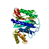
| ||||||||
|---|---|---|---|---|---|---|---|---|---|
| 1 |
| ||||||||
| Unit cell |
|
-Components
| #1: Protein | Mass: 32888.383 Da / Num. of mol.: 1 Source method: isolated from a genetically manipulated source Source: (gene. exp.) Pyrococcus furiosus (strain ATCC 43587 / DSM 3638 / JCM 8422 / Vc1) (archaea)Strain: ATCC 43587 / DSM 3638 / JCM 8422 / Vc1 / Gene: map, PF0541 / Production host:  | ||
|---|---|---|---|
| #2: Chemical |   Mass: 58.933 Da / Num. of mol.: 2 / Source method: obtained synthetically / Formula: Co / Feature type: SUBJECT OF INVESTIGATION Mass: 58.933 Da / Num. of mol.: 2 / Source method: obtained synthetically / Formula: Co / Feature type: SUBJECT OF INVESTIGATIONHas ligand of interest | Y | |
-Experimental details
-Experiment
| Experiment | Method: X-RAY DIFFRACTION / Number of used crystals: 1 |
|---|
- Sample preparation
| Crystal | Density Matthews: 5.1 Å3/Da / Density % sol: 75.9 % |
|---|---|
| Crystal grow | Temperature: 298 K / Method: vapor diffusion, hanging drop / pH: 4.5 / Details: 0.1 M sodium acetate, 2M NaCl , 5% glycerol |
-Data collection
| Diffraction | Mean temperature: 100 K / Serial crystal experiment: N | ||||||||||||||||||||||||||||||
|---|---|---|---|---|---|---|---|---|---|---|---|---|---|---|---|---|---|---|---|---|---|---|---|---|---|---|---|---|---|---|---|
| Diffraction source | Source: SYNCHROTRON / Site: RRCAT INDUS-2 / Beamline: PX-BL21 / Wavelength: 0.97949 Å | ||||||||||||||||||||||||||||||
| Detector | Type: MAR scanner 345 mm plate / Detector: IMAGE PLATE / Date: Jan 9, 2020 | ||||||||||||||||||||||||||||||
| Radiation | Protocol: SINGLE WAVELENGTH / Monochromatic (M) / Laue (L): M / Scattering type: x-ray | ||||||||||||||||||||||||||||||
| Radiation wavelength | Wavelength: 0.97949 Å / Relative weight: 1 | ||||||||||||||||||||||||||||||
| Reflection | Resolution: 2.85→45.86 Å / Num. obs: 14342 / % possible obs: 92.7 % / Redundancy: 6 % / CC1/2: 0.992 / Rmerge(I) obs: 0.181 / Rpim(I) all: 0.074 / Rrim(I) all: 0.196 / Net I/σ(I): 6.7 | ||||||||||||||||||||||||||||||
| Reflection shell | Diffraction-ID: 1
|
-Phasing
| Phasing | Method: molecular replacement | ||||||
|---|---|---|---|---|---|---|---|
| Phasing MR | R rigid body: 0.414
|
- Processing
| Software |
| ||||||||||||||||||||||||||||||||||||||||||||||||||||||||||||
|---|---|---|---|---|---|---|---|---|---|---|---|---|---|---|---|---|---|---|---|---|---|---|---|---|---|---|---|---|---|---|---|---|---|---|---|---|---|---|---|---|---|---|---|---|---|---|---|---|---|---|---|---|---|---|---|---|---|---|---|---|---|
| Refinement | Method to determine structure: MOLECULAR REPLACEMENT Starting model: 1WKM Resolution: 3.2→45.79 Å / Cor.coef. Fo:Fc: 0.927 / Cor.coef. Fo:Fc free: 0.903 / SU B: 24.705 / SU ML: 0.373 / Cross valid method: THROUGHOUT / σ(F): 0 / ESU R: 0.726 / ESU R Free: 0.448 Details: HYDROGENS HAVE BEEN ADDED IN THE RIDING POSITIONS U VALUES : REFINED INDIVIDUALLY
| ||||||||||||||||||||||||||||||||||||||||||||||||||||||||||||
| Solvent computation | Ion probe radii: 0.8 Å / Shrinkage radii: 0.8 Å / VDW probe radii: 1.2 Å | ||||||||||||||||||||||||||||||||||||||||||||||||||||||||||||
| Displacement parameters | Biso max: 99.21 Å2 / Biso mean: 55.423 Å2 / Biso min: 37.03 Å2
| ||||||||||||||||||||||||||||||||||||||||||||||||||||||||||||
| Refinement step | Cycle: final / Resolution: 3.2→45.79 Å
| ||||||||||||||||||||||||||||||||||||||||||||||||||||||||||||
| Refine LS restraints |
| ||||||||||||||||||||||||||||||||||||||||||||||||||||||||||||
| LS refinement shell | Resolution: 3.2→3.282 Å / Rfactor Rfree error: 0
|
