 Movie
Movie Controller

[English] 日本語
 Yorodumi
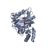
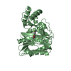
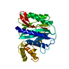
- PDB-1xgn: METHIONINE AMINOPEPTIDASE FROM HYPERTHERMOPHILE PYROCOCCUS FURIOSUS -
+ Open data

- Basic information
| Entry | Database: PDB / ID: 1xgn | ||||||
|---|---|---|---|---|---|---|---|
| Title | METHIONINE AMINOPEPTIDASE FROM HYPERTHERMOPHILE PYROCOCCUS FURIOSUS | ||||||
 Components Components | METHIONINE AMINOPEPTIDASE | ||||||
 Keywords Keywords | AMINOPEPTIDASE / HYPERTHERMOPHILE | ||||||
| Function / homology |  Function and homology information Function and homology informationmethionyl aminopeptidase / initiator methionyl aminopeptidase activity / metalloaminopeptidase activity / proteolysis / metal ion binding / cytoplasm Similarity search - Function | ||||||
| Biological species |   Pyrococcus furiosus (archaea) Pyrococcus furiosus (archaea) | ||||||
| Method |  X-RAY DIFFRACTION / MOLECULAR REPLACEMENT / Resolution: 2.9 Å X-RAY DIFFRACTION / MOLECULAR REPLACEMENT / Resolution: 2.9 Å | ||||||
 Authors Authors | Tahirov, T.H. / Tsukihara, T. | ||||||
 Citation Citation | Journal: J.Mol.Biol. / Year: 1998 Title: Crystal structure of methionine aminopeptidase from hyperthermophile, Pyrococcus furiosus. Authors: Tahirov, T.H. / Oki, H. / Tsukihara, T. / Ogasahara, K. / Yutani, K. / Ogata, K. / Izu, Y. / Tsunasawa, S. / Kato, I. #1: Journal: Acta Crystallogr.,Sect.D / Year: 1997Title: Crystallization and Preliminary X-Ray Analysis of Methionine Aminopeptidase from the Hyperthermophilic Bacterium Pyrococcus Furiosus Authors: Tahirov, T.H. / Oki, H. / Tsukihara, T. / Ogasahara, K. / Izu, Y. / Tsunasawa, S. / Kato, I. / Yutani, K. | ||||||
| History |
|
- Structure visualization
| Structure viewer | Molecule: MolmilJmol/JSmol |
|---|
- Downloads & links
-Download
| PDBx/mmCIF format | 1xgn.cif.gz | 124.6 KB | Display | PDBx/mmCIF format |
|---|---|---|---|---|
| PDB format | pdb1xgn.ent.gz | 98.9 KB | Display | PDB format |
| PDBx/mmJSON format | 1xgn.json.gz | Tree view | PDBx/mmJSON format | |
| Others |  Other downloads Other downloads |
-Validation report
| Arichive directory | https://data.pdbj.org/pub/pdb/validation_reports/xg/1xgnftp://data.pdbj.org/pub/pdb/validation_reports/xg/1xgn | HTTPS FTP |
|---|
-Related structure data
| Related structure data |  1xgmSC  1xgoC 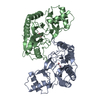 1xgsC S: Starting model for refinement C: citing same article ( |
|---|---|
| Similar structure data |


-Links
 PDBj
PDBj
- Assembly
| Deposited unit | 
| ||||||||
|---|---|---|---|---|---|---|---|---|---|
| 1 | 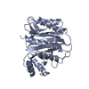
| ||||||||
| 2 | 
| ||||||||
| Unit cell |
| ||||||||
| Noncrystallographic symmetry (NCS) | NCS oper: (Code: given Matrix: (-0.451342, 0.868787, -0.203715), Vector: |
-Components
| #1: Protein | Mass: 32888.383 Da / Num. of mol.: 2 / Source method: isolated from a natural source / Details: TWO COBALT IONS IN ACTIVE SITE / Source: (natural) Pyrococcus furiosus (archaea) / References: UniProt: P56218, methionyl aminopeptidase#2: Chemical | ChemComp-CO /   Mass: 58.933 Da / Num. of mol.: 4 / Source method: obtained synthetically / Formula: Co Mass: 58.933 Da / Num. of mol.: 4 / Source method: obtained synthetically / Formula: Co#3: Water | ChemComp-HOH / |  Mass: 18.015 Da / Num. of mol.: 25 / Source method: isolated from a natural source / Formula: H2O Mass: 18.015 Da / Num. of mol.: 25 / Source method: isolated from a natural source / Formula: H2O |
|---|
-Experimental details
-Experiment
| Experiment | Method: X-RAY DIFFRACTION / Number of used crystals: 1 |
|---|
- Sample preparation
| Crystal | Density Matthews: 3.38 Å3/Da / Density % sol: 63.7 % | ||||||||||||||||||||||||||||||||||||||||||
|---|---|---|---|---|---|---|---|---|---|---|---|---|---|---|---|---|---|---|---|---|---|---|---|---|---|---|---|---|---|---|---|---|---|---|---|---|---|---|---|---|---|---|---|
| Crystal grow | pH: 7.5 Details: PROTEIN SOLUTION CONTAINING 16 MG/ML PFMAP, 2 MM COCL2 AND 30 MM L-METHIONINE IN 20 MM POTASSIUM ACETATE AT PH4.5 WAS MIXED WITH EQUAL AMOUNT OF RESERVOIR SOLUTION CONTAINING 0.6% PROPANOL ...Details: PROTEIN SOLUTION CONTAINING 16 MG/ML PFMAP, 2 MM COCL2 AND 30 MM L-METHIONINE IN 20 MM POTASSIUM ACETATE AT PH4.5 WAS MIXED WITH EQUAL AMOUNT OF RESERVOIR SOLUTION CONTAINING 0.6% PROPANOL AND 1.6% PEG400 IN 0.1 M SODIUM HEPES BUFFER AT PH 7.5, THEN EQUILIBRATED AGAINST RESERVOIR SOLUTION. PH range: 4.5-7.5 | ||||||||||||||||||||||||||||||||||||||||||
| Crystal | *PLUS | ||||||||||||||||||||||||||||||||||||||||||
| Crystal grow | *PLUS Temperature: 277 K / pH: 4.5 / Method: vapor diffusion, sitting dropDetails: Tahirov, T.H., (1997) Acta Crystallogr.,Sect.D, 53, 798. | ||||||||||||||||||||||||||||||||||||||||||
| Components of the solutions | *PLUS
|
-Data collection
| Diffraction | Mean temperature: 289 K |
|---|---|
| Diffraction source | Source: ROTATING ANODE / Type: RIGAKU RUH2R / Wavelength: 1.5418 |
| Detector | Type: RIGAKU / Detector: IMAGE PLATE / Date: Jun 1, 1996 |
| Radiation | Monochromator: GRAPHITE(002) / Monochromatic (M) / Laue (L): M / Scattering type: x-ray |
| Radiation wavelength | Wavelength: 1.5418 Å / Relative weight: 1 |
| Reflection | Resolution: 2.9→50 Å / Num. obs: 17502 / % possible obs: 90 % / Observed criterion σ(I): 1 / Redundancy: 2.81 % / Rmerge(I) obs: 0.072 / Net I/σ(I): 12.4 |
| Reflection shell | Resolution: 2.9→3.03 Å / Rmerge(I) obs: 0.245 / Mean I/σ(I) obs: 2.1 / % possible all: 62.7 |
| Reflection | *PLUS Num. measured all: 91220 |
| Reflection shell | *PLUS % possible obs: 62.7 % |
- Processing
| Software |
| ||||||||||||||||||||||||||||||||||||||||||||||||||||||||||||
|---|---|---|---|---|---|---|---|---|---|---|---|---|---|---|---|---|---|---|---|---|---|---|---|---|---|---|---|---|---|---|---|---|---|---|---|---|---|---|---|---|---|---|---|---|---|---|---|---|---|---|---|---|---|---|---|---|---|---|---|---|---|
| Refinement | Method to determine structure: MOLECULAR REPLACEMENT Starting model: MONOCLINIC CRYSTAL FORM, PDB ENTRY 1XGM Resolution: 2.9→15 Å / Data cutoff high absF: 1000000 / Data cutoff low absF: 0.001 / Cross valid method: THROUGHOUT / σ(F): 2 Details: PARAMETER AND TOPOLOGY FILES ARE MODIFIED TO INCLUDE THE COBALT IONS IN THE REFINEMENT.
| ||||||||||||||||||||||||||||||||||||||||||||||||||||||||||||
| Displacement parameters | Biso mean: 37.4 Å2 | ||||||||||||||||||||||||||||||||||||||||||||||||||||||||||||
| Refine analyze | Luzzati coordinate error obs: 0.26 Å | ||||||||||||||||||||||||||||||||||||||||||||||||||||||||||||
| Refinement step | Cycle: LAST / Resolution: 2.9→15 Å
| ||||||||||||||||||||||||||||||||||||||||||||||||||||||||||||
| Refine LS restraints |
| ||||||||||||||||||||||||||||||||||||||||||||||||||||||||||||
| LS refinement shell | Resolution: 2.9→3.03 Å / Total num. of bins used: 8
| ||||||||||||||||||||||||||||||||||||||||||||||||||||||||||||
| Xplor file |
| ||||||||||||||||||||||||||||||||||||||||||||||||||||||||||||
| Software | *PLUS Name: X-PLOR / Version: 3.1 / Classification: refinement | ||||||||||||||||||||||||||||||||||||||||||||||||||||||||||||
| Refine LS restraints | *PLUS
| ||||||||||||||||||||||||||||||||||||||||||||||||||||||||||||
| LS refinement shell | *PLUS Rfactor Rfree: 0.34 |
