 Movie
Movie Controller


+ Open data

- Basic information
| Entry | Database: PDB / ID: 6lyf | ||||||
|---|---|---|---|---|---|---|---|
| Title | Crystal structure of the mouse endonuclease EndoG(H138A/Se-Met) | ||||||
 Components Components | Endonuclease G, mitochondrial | ||||||
 Keywords Keywords | APOPTOSIS / EndoG / DNase / mitochondria | ||||||
| Function / homology |  Function and homology information Function and homology informationmitochondrial DNA catabolic process / positive regulation of hydrogen peroxide-mediated programmed cell death / Hydrolases; Acting on ester bonds; Endoribonucleases that are active with either ribo- or deoxyribonucleic acids and produce 5'-phosphomonoesters / positive regulation of mitochondrial DNA replication / DNA nuclease activity / positive regulation of apoptotic DNA fragmentation / single-stranded DNA endonuclease activity / apoptotic DNA fragmentation / negative regulation of TOR signaling / DNA catabolic process ...mitochondrial DNA catabolic process / positive regulation of hydrogen peroxide-mediated programmed cell death / Hydrolases; Acting on ester bonds; Endoribonucleases that are active with either ribo- or deoxyribonucleic acids and produce 5'-phosphomonoesters / positive regulation of mitochondrial DNA replication / DNA nuclease activity / positive regulation of apoptotic DNA fragmentation / single-stranded DNA endonuclease activity / apoptotic DNA fragmentation / negative regulation of TOR signaling / DNA catabolic process / response to tumor necrosis factor / response to mechanical stimulus / RNA endonuclease activity / positive regulation of autophagy / cellular response to calcium ion / DNA endonuclease activity / cellular response to glucose stimulus / response to estradiol / cellular response to oxidative stress / cellular response to hypoxia / nucleic acid binding / in utero embryonic development / perikaryon / mitochondrial inner membrane / positive regulation of apoptotic process / response to antibiotic / DNA damage response / perinuclear region of cytoplasm / magnesium ion binding / protein homodimerization activity / mitochondrion / nucleus Similarity search - Function | ||||||
| Biological species |  | ||||||
| Method |  X-RAY DIFFRACTION / SYNCHROTRON / SAD / Resolution: 2.8 Å X-RAY DIFFRACTION / SYNCHROTRON / SAD / Resolution: 2.8 Å | ||||||
 Authors Authors | Park, K.H. / Woo, E.J. | ||||||
 Citation Citation | Journal: Biochem.Biophys.Res.Commun. / Year: 2020 Title: Crystal structure of the mouse endonuclease G. Authors: Park, K.H. / Yoon, S.M. / Song, H.N. / Yang, J.H. / Ryu, S.E. / Woo, E.J. | ||||||
| History |
|
- Structure visualization
| Structure viewer | Molecule: MolmilJmol/JSmol |
|---|
- Downloads & links
-Download
| PDBx/mmCIF format | 6lyf.cif.gz | 440.7 KB | Display | PDBx/mmCIF format |
|---|---|---|---|---|
| PDB format | pdb6lyf.ent.gz | 304.2 KB | Display | PDB format |
| PDBx/mmJSON format | 6lyf.json.gz | Tree view | PDBx/mmJSON format | |
| Others |  Other downloads Other downloads |
-Validation report
| Arichive directory | https://data.pdbj.org/pub/pdb/validation_reports/ly/6lyfftp://data.pdbj.org/pub/pdb/validation_reports/ly/6lyf | HTTPS FTP |
|---|
-Related structure data


-Links
 PDBj
PDBj- Assembly
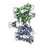
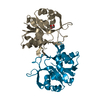
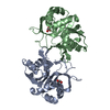
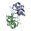
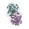
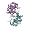
| Deposited unit | 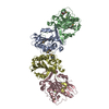
| ||||||||||||
|---|---|---|---|---|---|---|---|---|---|---|---|---|---|
| 1 | 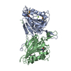
| ||||||||||||
| 2 | 
| ||||||||||||
| Unit cell |
| ||||||||||||
| Noncrystallographic symmetry (NCS) | NCS domain:
|
-Components
| #1: Protein | Mass: 27598.734 Da / Num. of mol.: 4 / Mutation: H138A Source method: isolated from a genetically manipulated source Source: (gene. exp.)  References: UniProt: O08600, Hydrolases; Acting on ester bonds; Endoribonucleases that are active with either ribo- or deoxyribonucleic acids and produce 5'-phosphomonoesters #2: Chemical | ChemComp-MG /   Mass: 24.305 Da / Num. of mol.: 4 / Source method: obtained synthetically / Formula: Mg / Feature type: SUBJECT OF INVESTIGATION Mass: 24.305 Da / Num. of mol.: 4 / Source method: obtained synthetically / Formula: Mg / Feature type: SUBJECT OF INVESTIGATION#3: Water | ChemComp-HOH / |  Mass: 18.015 Da / Num. of mol.: 180 / Source method: isolated from a natural source / Formula: H2O Mass: 18.015 Da / Num. of mol.: 180 / Source method: isolated from a natural source / Formula: H2OHas ligand of interest | Y | Has protein modification | Y | |
|---|
-Experimental details
-Experiment
| Experiment | Method: X-RAY DIFFRACTION / Number of used crystals: 1 |
|---|
- Sample preparation
| Crystal | Density Matthews: 2.35 Å3/Da / Density % sol: 47.73 % |
|---|---|
| Crystal grow | Temperature: 291 K / Method: vapor diffusion, sitting drop Details: 200mM NaCl, 1mM CdCl2, 100mM CHES, 10mM CTAB, 5% glycerol, 5% MPD |
-Data collection
| Diffraction | Mean temperature: 193 K / Serial crystal experiment: N |
|---|---|
| Diffraction source | Source: SYNCHROTRON / Site: EMBL/DESY, HAMBURG  / Beamline: BW7A / Wavelength: 1 Å / Beamline: BW7A / Wavelength: 1 Å |
| Detector | Type: ADSC QUANTUM 270 / Detector: CCD / Date: Apr 13, 2010 |
| Radiation | Protocol: SINGLE WAVELENGTH / Monochromatic (M) / Laue (L): M / Scattering type: x-ray |
| Radiation wavelength | Wavelength: 1 Å / Relative weight: 1 |
| Reflection | Resolution: 2.8→48.12 Å / Num. obs: 56219 / % possible obs: 99.7 % / Redundancy: 14.3 % / Biso Wilson estimate: 39.44 Å2 / Rsym value: 0.086 / Net I/σ(I): 13.5 |
| Reflection shell | Resolution: 2.8→2.85 Å / Num. unique obs: 28825 / Rsym value: 0.4 |
- Processing
| Software |
| ||||||||||||||||||||||||||||||||||||||||||||||||||||||||||||||||||||||||||||||||||||||||||||||||||||||||||||||||||||||||||||||
|---|---|---|---|---|---|---|---|---|---|---|---|---|---|---|---|---|---|---|---|---|---|---|---|---|---|---|---|---|---|---|---|---|---|---|---|---|---|---|---|---|---|---|---|---|---|---|---|---|---|---|---|---|---|---|---|---|---|---|---|---|---|---|---|---|---|---|---|---|---|---|---|---|---|---|---|---|---|---|---|---|---|---|---|---|---|---|---|---|---|---|---|---|---|---|---|---|---|---|---|---|---|---|---|---|---|---|---|---|---|---|---|---|---|---|---|---|---|---|---|---|---|---|---|---|---|---|---|
| Refinement | Method to determine structure: SAD / Resolution: 2.8→48.12 Å / SU ML: 0.3627 / Cross valid method: FREE R-VALUE / σ(F): 1.36 / Phase error: 26.2075
| ||||||||||||||||||||||||||||||||||||||||||||||||||||||||||||||||||||||||||||||||||||||||||||||||||||||||||||||||||||||||||||||
| Solvent computation | Shrinkage radii: 0.9 Å / VDW probe radii: 1.11 Å | ||||||||||||||||||||||||||||||||||||||||||||||||||||||||||||||||||||||||||||||||||||||||||||||||||||||||||||||||||||||||||||||
| Displacement parameters | Biso mean: 49.19 Å2 | ||||||||||||||||||||||||||||||||||||||||||||||||||||||||||||||||||||||||||||||||||||||||||||||||||||||||||||||||||||||||||||||
| Refinement step | Cycle: LAST / Resolution: 2.8→48.12 Å
| ||||||||||||||||||||||||||||||||||||||||||||||||||||||||||||||||||||||||||||||||||||||||||||||||||||||||||||||||||||||||||||||
| Refine LS restraints |
| ||||||||||||||||||||||||||||||||||||||||||||||||||||||||||||||||||||||||||||||||||||||||||||||||||||||||||||||||||||||||||||||
| LS refinement shell |
| ||||||||||||||||||||||||||||||||||||||||||||||||||||||||||||||||||||||||||||||||||||||||||||||||||||||||||||||||||||||||||||||
| Refinement TLS params. | Method: refined / Origin x: 10.3241861153 Å / Origin y: 27.5101005381 Å / Origin z: 67.3070337233 Å
| ||||||||||||||||||||||||||||||||||||||||||||||||||||||||||||||||||||||||||||||||||||||||||||||||||||||||||||||||||||||||||||||
| Refinement TLS group | Selection details: all |
