- PDB-6m3f: Crystal structure of the mouse endonuclease EndoG(H138A/C110A), s... -
+
Open data
ID or keywords:
Loading...
-
Basic information
Entry
Database: PDB / ID: 6m3f
Title
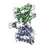
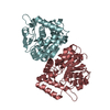
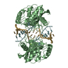
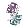
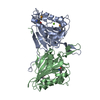
Crystal structure of the mouse endonuclease EndoG(H138A/C110A), space group P212121
Components
Endonuclease G, mitochondrial
Keywords
APOPTOSIS / EndoG / DNase / mitochondria
Function / homology
Function and homology information
mitochondrial DNA catabolic process / positive regulation of hydrogen peroxide-mediated programmed cell death / Hydrolases; Acting on ester bonds; Endoribonucleases that are active with either ribo- or deoxyribonucleic acids and produce 5'-phosphomonoesters / positive regulation of mitochondrial DNA replication / DNA nuclease activity / positive regulation of apoptotic DNA fragmentation / single-stranded DNA endonuclease activity / apoptotic DNA fragmentation / negative regulation of TOR signaling / DNA catabolic process ...mitochondrial DNA catabolic process / positive regulation of hydrogen peroxide-mediated programmed cell death / Hydrolases; Acting on ester bonds; Endoribonucleases that are active with either ribo- or deoxyribonucleic acids and produce 5'-phosphomonoesters / positive regulation of mitochondrial DNA replication / DNA nuclease activity / positive regulation of apoptotic DNA fragmentation / single-stranded DNA endonuclease activity / apoptotic DNA fragmentation / negative regulation of TOR signaling / DNA catabolic process / response to tumor necrosis factor / response to mechanical stimulus / RNA endonuclease activity / positive regulation of autophagy / cellular response to calcium ion / DNA endonuclease activity / cellular response to glucose stimulus / response to estradiol / cellular response to oxidative stress / cellular response to hypoxia / nucleic acid binding / in utero embryonic development / perikaryon / mitochondrial inner membrane / positive regulation of apoptotic process / response to antibiotic / DNA damage response / perinuclear region of cytoplasm / magnesium ion binding / protein homodimerization activity / mitochondrion / nucleus Similarity search - Function
DNA/RNA non-specific endonuclease, active site / DNA/RNA non-specific endonucleases active site. / Non-specific endonuclease / Extracellular Endonuclease; Chain A / Extracellular Endonuclease, subunit A / Extracellular Endonuclease, subunit A / DNA/RNA non-specific endonuclease / DNA/RNA non-specific endonuclease / DNA/RNA non-specific endonuclease / DNA/RNA non-specific endonuclease ...DNA/RNA non-specific endonuclease, active site / DNA/RNA non-specific endonucleases active site. / Non-specific endonuclease / Extracellular Endonuclease; Chain A / Extracellular Endonuclease, subunit A / Extracellular Endonuclease, subunit A / DNA/RNA non-specific endonuclease / DNA/RNA non-specific endonuclease / DNA/RNA non-specific endonuclease / DNA/RNA non-specific endonuclease / DNA/RNA non-specific endonuclease superfamily / His-Me finger superfamily / 3-Layer(aba) Sandwich / Alpha Beta Similarity search - Domain/homology
Mass: 27472.879 Da / Num. of mol.: 2 / Mutation: H138A/C110A Source method: isolated from a genetically manipulated source Source: (gene. exp.) Mus musculus (house mouse) / Gene: Endog / Production host: Escherichia coli (E. coli) References: UniProt: O08600, Hydrolases; Acting on ester bonds; Endoribonucleases that are active with either ribo- or deoxyribonucleic acids and produce 5'-phosphomonoesters
In the structure databanks used in Yorodumi, some data are registered as the other names, "COVID-19 virus" and "2019-nCoV". Here are the details of the virus and the list of structure data.
Jan 31, 2019. EMDB accession codes are about to change! (news from PDBe EMDB page)
EMDB accession codes are about to change! (news from PDBe EMDB page)
The allocation of 4 digits for EMDB accession codes will soon come to an end. Whilst these codes will remain in use, new EMDB accession codes will include an additional digit and will expand incrementally as the available range of codes is exhausted. The current 4-digit format prefixed with “EMD-” (i.e. EMD-XXXX) will advance to a 5-digit format (i.e. EMD-XXXXX), and so on. It is currently estimated that the 4-digit codes will be depleted around Spring 2019, at which point the 5-digit format will come into force.
The EM Navigator/Yorodumi systems omit the EMD- prefix.
Related info.:Q: What is EMD? / ID/Accession-code notation in Yorodumi/EM Navigator
Yorodumi is a browser for structure data from EMDB, PDB, SASBDB, etc.
This page is also the successor to EM Navigator detail page, and also detail information page/front-end page for Omokage search.
The word "yorodu" (or yorozu) is an old Japanese word meaning "ten thousand". "mi" (miru) is to see.
Related info.:EMDB / PDB / SASBDB / Comparison of 3 databanks / Yorodumi Search / Aug 31, 2016. New EM Navigator & Yorodumi / Yorodumi Papers / Jmol/JSmol / Function and homology information / Changes in new EM Navigator and Yorodumi
 Movie
Movie Controller

 Yorodumi
 Open data
 Basic information
 Components
 Keywords
 Function and homology information
 X-RAY DIFFRACTION /
  Authors
 Citation
 Structure visualization
 Downloads & links
 Other downloads


 PDBj
PDBj Assembly


 Mass: 24.305 Da / Num. of mol.: 2 / Source method: obtained synthetically / Formula: Mg / Feature type: SUBJECT OF INVESTIGATION
 Mass: 18.015 Da / Num. of mol.: 300 / Source method: isolated from a natural source / Formula: H2O
 Sample preparation
 / Beamline: 6D / Wavelength: 1 Å
 Processing