 Movie
Movie Controller

[English] 日本語
 Yorodumi
- PDB-6lxw: Cryo-EM structure of human secretory immunoglobulin A in complex ... -
+ Open data

- Basic information
| Entry | Database: PDB / ID: 6lxw | |||||||||||||||||||||||||||||||||||||||||||||||||||||||||||||||
|---|---|---|---|---|---|---|---|---|---|---|---|---|---|---|---|---|---|---|---|---|---|---|---|---|---|---|---|---|---|---|---|---|---|---|---|---|---|---|---|---|---|---|---|---|---|---|---|---|---|---|---|---|---|---|---|---|---|---|---|---|---|---|---|---|
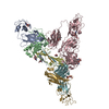
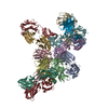
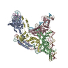
| Title | Cryo-EM structure of human secretory immunoglobulin A in complex with the N-terminal domain of SpsA | |||||||||||||||||||||||||||||||||||||||||||||||||||||||||||||||
 Components Components |
| |||||||||||||||||||||||||||||||||||||||||||||||||||||||||||||||
 Keywords Keywords | IMMUNE SYSTEM / immunoglobulin / dimer / transcytosis / secreted | |||||||||||||||||||||||||||||||||||||||||||||||||||||||||||||||
| Function / homology |  Function and homology information Function and homology informationpolymeric immunoglobulin receptor activity / immunoglobulin transcytosis in epithelial cells mediated by polymeric immunoglobulin receptor / polymeric immunoglobulin binding / response to tacrolimus / kappa-type opioid receptor binding / regulation of CD4-positive, alpha-beta T cell proliferation / regulation of T cell homeostatic proliferation / dimeric IgA immunoglobulin complex / interleukin-2 receptor binding / secretory dimeric IgA immunoglobulin complex ...polymeric immunoglobulin receptor activity / immunoglobulin transcytosis in epithelial cells mediated by polymeric immunoglobulin receptor / polymeric immunoglobulin binding / response to tacrolimus / kappa-type opioid receptor binding / regulation of CD4-positive, alpha-beta T cell proliferation / regulation of T cell homeostatic proliferation / dimeric IgA immunoglobulin complex / interleukin-2 receptor binding / secretory dimeric IgA immunoglobulin complex / monomeric IgA immunoglobulin complex / pentameric IgM immunoglobulin complex / secretory IgA immunoglobulin complex / negative regulation of lymphocyte proliferation / glycosphingolipid binding / positive regulation of plasma cell differentiation / Fc receptor signaling pathway / positive regulation of tissue remodeling / IgA binding / negative regulation of T-helper 17 cell differentiation / IgA immunoglobulin complex / RUNX1 and FOXP3 control the development of regulatory T lymphocytes (Tregs) / glomerular filtration / leukocyte activation involved in immune response / positive regulation of isotype switching to IgG isotypes / detection of chemical stimulus involved in sensory perception of bitter taste / activated T cell proliferation / Interleukin-2 signaling / cell surface receptor signaling pathway via STAT / natural killer cell activation / kinase activator activity / positive regulation of activated T cell proliferation / immunoglobulin complex, circulating / positive regulation of regulatory T cell differentiation / negative regulation of B cell apoptotic process / immunoglobulin receptor binding / azurophil granule membrane / IgG immunoglobulin complex / interleukin-2-mediated signaling pathway / positive regulation of immunoglobulin production / positive regulation of interleukin-17 production / positive regulation of dendritic spine development / positive regulation of respiratory burst / receptor clustering / humoral immune response / T cell differentiation / complement activation, classical pathway / Interleukin receptor SHC signaling / Scavenging of heme from plasma / antigen binding / extrinsic apoptotic signaling pathway in absence of ligand / positive regulation of B cell proliferation / cytokine activity / Cell surface interactions at the vascular wall / B cell receptor signaling pathway / growth factor activity / negative regulation of inflammatory response / positive regulation of type II interferon production / epidermal growth factor receptor signaling pathway / positive regulation of inflammatory response / transmembrane signaling receptor activity / cell-cell signaling / antibacterial humoral response / carbohydrate binding / positive regulation of cytosolic calcium ion concentration / RAF/MAP kinase cascade / positive regulation of cell growth / protein-containing complex assembly / blood microparticle / transcription by RNA polymerase II / phospholipase C-activating G protein-coupled receptor signaling pathway / adaptive immune response / response to ethanol / protein-macromolecule adaptor activity / cell adhesion / signaling receptor complex / immune response / innate immune response / positive regulation of cell population proliferation / Neutrophil degranulation / negative regulation of apoptotic process / signal transduction / protein homodimerization activity / positive regulation of transcription by RNA polymerase II / : / extracellular exosome / extracellular region / plasma membrane Similarity search - Function | |||||||||||||||||||||||||||||||||||||||||||||||||||||||||||||||
| Biological species |  Homo sapiens (human) Homo sapiens (human)  Streptococcus pneumoniae (bacteria) Streptococcus pneumoniae (bacteria) | |||||||||||||||||||||||||||||||||||||||||||||||||||||||||||||||
| Method | ELECTRON MICROSCOPY / single particle reconstruction / cryo EM / Resolution: 3.27 Å | |||||||||||||||||||||||||||||||||||||||||||||||||||||||||||||||
 Authors Authors | Wang, Y. / Wang, G. / Li, Y. / Xiao, J. | |||||||||||||||||||||||||||||||||||||||||||||||||||||||||||||||
| Funding support |  China, 1items China, 1items
| |||||||||||||||||||||||||||||||||||||||||||||||||||||||||||||||
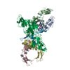
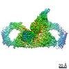
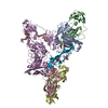
 Citation Citation | Journal: Cell Res / Year: 2020 Title: Structural insights into secretory immunoglobulin A and its interaction with a pneumococcal adhesin. Authors: Yuxin Wang / Guopeng Wang / Yaxin Li / Qinyu Zhu / Hao Shen / Ning Gao / Junyu Xiao / Abstract: Secretory Immunoglobulin A (SIgA) is the most abundant antibody at the mucosal surface. It possesses two additional subunits besides IgA: the joining chain (J-chain) and secretory component (SC). SC ...Secretory Immunoglobulin A (SIgA) is the most abundant antibody at the mucosal surface. It possesses two additional subunits besides IgA: the joining chain (J-chain) and secretory component (SC). SC is the ectodomain of the polymeric immunoglobulin receptor (pIgR), which functions to transport IgA to the mucosa. How the J-chain and pIgR/SC facilitate the assembly and secretion of SIgA remains incompletely understood. Furthermore, during the infection of Streptococcus pneumoniae, the pneumococcal adhesin SpsA hijacks pIgR/SC and SIgA to gain entry to human cells and evade host defense. How SpsA targets pIgR/SC and SIgA also remains elusive. Here we report a cryo-electron microscopy structure of the Fc region of IgA1 (Fcα) in complex with the J-chain and SC (Fcα-J-SC), which reveals the organization principle of SIgA. We also present a structure of Fcα-J-SC complexed with SpsA, which uncovers the specific interactions between SpsA and human pIgR/SC. These results advance the molecular understanding of SIgA and shed light on S. pneumoniae pathogenesis. | |||||||||||||||||||||||||||||||||||||||||||||||||||||||||||||||
| History |
|
- Structure visualization
| Movie |
Movie viewer |
|---|---|
| Structure viewer | Molecule: MolmilJmol/JSmol |
- Downloads & links
-Download
| PDBx/mmCIF format | 6lxw.cif.gz | 294.1 KB | Display | PDBx/mmCIF format |
|---|---|---|---|---|
| PDB format | pdb6lxw.ent.gz | 228.6 KB | Display | PDB format |
| PDBx/mmJSON format | 6lxw.json.gz | Tree view | PDBx/mmJSON format | |
| Others |  Other downloads Other downloads |
-Validation report
| Arichive directory | https://data.pdbj.org/pub/pdb/validation_reports/lx/6lxwftp://data.pdbj.org/pub/pdb/validation_reports/lx/6lxw | HTTPS FTP |
|---|
-Related structure data
| Related structure data |  30008MC  6lx3C M: map data used to model this data C: citing same article ( |
|---|---|
| Similar structure data |


-Links
 PDBj
PDBj


- Assembly
| Deposited unit | 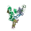
|
|---|---|
| 1 |
|
-Components
| #1: Antibody | Mass: 31445.693 Da / Num. of mol.: 4 Source method: isolated from a genetically manipulated source Source: (gene. exp.) Homo sapiens (human) / Gene: IL2, IGHA1 / Production host: Homo sapiens (human) / References: UniProt: P60568, UniProt: P01876#2: Protein | | Mass: 19225.762 Da / Num. of mol.: 1 Source method: isolated from a genetically manipulated source Source: (gene. exp.) Homo sapiens (human) / Gene: JCHAIN, IGCJ, IGJ / Production host: Homo sapiens (human) / References: UniProt: P01591#3: Protein | | Mass: 63306.336 Da / Num. of mol.: 1 Source method: isolated from a genetically manipulated source Source: (gene. exp.) Homo sapiens (human) / Gene: PIGR / Production host: Homo sapiens (human) / References: UniProt: P01833#4: Protein | | Mass: 36311.113 Da / Num. of mol.: 1 Source method: isolated from a genetically manipulated source Source: (gene. exp.) Streptococcus pneumoniae (bacteria) / Gene: spsA / Production host:  Escherichia phage Ecwhy_1 (virus) / References: UniProt: O33753 Escherichia phage Ecwhy_1 (virus) / References: UniProt: O33753Has protein modification | Y | |
|---|
-Experimental details
-Experiment
| Experiment | Method: ELECTRON MICROSCOPY |
|---|---|
| EM experiment | Aggregation state: PARTICLE / 3D reconstruction method: single particle reconstruction |
- Sample preparation
| Component | Name: Quadruple complex of human secretory immunoglobulin A with the N-terminal domain of SpsA Type: COMPLEX / Entity ID: all / Source: RECOMBINANT |
|---|---|
| Source (natural) | Organism: Homo sapiens (human) |
| Source (recombinant) | Organism: Homo sapiens (human) |
| Buffer solution | pH: 7.4 |
| Specimen | Conc.: 0.2 mg/ml / Embedding applied: NO / Shadowing applied: NO / Staining applied: NO / Vitrification applied: YES |
| Vitrification | Cryogen name: ETHANE / Humidity: 100 % / Chamber temperature: 277.15 K |
- Electron microscopy imaging
| Experimental equipment |  Model: Titan Krios / Image courtesy: FEI Company |
|---|---|
| Microscopy | Model: FEI TITAN KRIOS |
| Electron gun | Electron source:  FIELD EMISSION GUN / Accelerating voltage: 300 kV / Illumination mode: FLOOD BEAM FIELD EMISSION GUN / Accelerating voltage: 300 kV / Illumination mode: FLOOD BEAM |
| Electron lens | Mode: BRIGHT FIELD |
| Image recording | Electron dose: 59.74 e/Å2 / Detector mode: SUPER-RESOLUTION / Film or detector model: GATAN K2 QUANTUM (4k x 4k) |
- Processing
| Software | Name: PHENIX / Version: 1.15.2_3472: / Classification: refinement | ||||||||||||||||||||||||
|---|---|---|---|---|---|---|---|---|---|---|---|---|---|---|---|---|---|---|---|---|---|---|---|---|---|
| EM software | Name: PHENIX / Category: model refinement | ||||||||||||||||||||||||
| CTF correction | Type: PHASE FLIPPING AND AMPLITUDE CORRECTION | ||||||||||||||||||||||||
| Symmetry | Point symmetry: C1 (asymmetric) | ||||||||||||||||||||||||
| 3D reconstruction | Resolution: 3.27 Å / Resolution method: FSC 0.143 CUT-OFF / Num. of particles: 280791 / Symmetry type: POINT | ||||||||||||||||||||||||
| Refine LS restraints |
|
