 Movie
Movie Controller

[English] 日本語
 Yorodumi
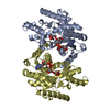
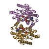
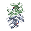
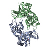
- PDB-6lut: Crystal structure of Serine Racemase from Dictyostelium discoideum. -
+ Open data

- Basic information
| Entry | Database: PDB / ID: 6lut | ||||||
|---|---|---|---|---|---|---|---|
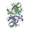
| Title | Crystal structure of Serine Racemase from Dictyostelium discoideum. | ||||||
 Components Components | Probable serine racemase | ||||||
 Keywords Keywords | ISOMERASE / D-amino acid / Racemase / PLP | ||||||
| Function / homology |  Function and homology information Function and homology informationprotein dehydration / Serine metabolism / D-serine biosynthetic process / serine racemase / threonine racemase activity / serine racemase activity / D-serine ammonia-lyase / L-serine ammonia-lyase / D-serine ammonia-lyase activity / L-serine ammonia-lyase activity ...protein dehydration / Serine metabolism / D-serine biosynthetic process / serine racemase / threonine racemase activity / serine racemase activity / D-serine ammonia-lyase / L-serine ammonia-lyase / D-serine ammonia-lyase activity / L-serine ammonia-lyase activity / D-serine catabolic process / pyruvate biosynthetic process / L-serine catabolic process / L-serine metabolic process / pyridoxal phosphate binding / magnesium ion binding / ATP binding / cytoplasm Similarity search - Function | ||||||
| Biological species |  | ||||||
| Method |  X-RAY DIFFRACTION / SYNCHROTRON / MOLECULAR REPLACEMENT / Resolution: 1.35 Å X-RAY DIFFRACTION / SYNCHROTRON / MOLECULAR REPLACEMENT / Resolution: 1.35 Å | ||||||
 Authors Authors | Goto, M. / Mizobuchi, T. / Yoshimura, T. | ||||||
 Citation Citation | Journal: Biochim Biophys Acta Proteins Proteom / Year: 2020 Title: Mechanism of eukaryotic serine racemase-catalyzed serine dehydration. Authors: Ito, T. / Matsuoka, M. / Goto, M. / Watanabe, S. / Mizobuchi, T. / Matsushita, K. / Nasu, R. / Hemmi, H. / Yoshimura, T. | ||||||
| History |
|
- Structure visualization
| Structure viewer | Molecule: MolmilJmol/JSmol |
|---|
- Downloads & links
-Download
| PDBx/mmCIF format | 6lut.cif.gz | 139.4 KB | Display | PDBx/mmCIF format |
|---|---|---|---|---|
| PDB format | pdb6lut.ent.gz | 104.7 KB | Display | PDB format |
| PDBx/mmJSON format | 6lut.json.gz | Tree view | PDBx/mmJSON format | |
| Others |  Other downloads Other downloads |
-Validation report
| Arichive directory | https://data.pdbj.org/pub/pdb/validation_reports/lu/6lutftp://data.pdbj.org/pub/pdb/validation_reports/lu/6lut | HTTPS FTP |
|---|
-Related structure data
| Related structure data |  1v71S S: Starting model for refinement |
|---|---|
| Similar structure data |

-Links
 PDBj
PDBj
- Assembly
| Deposited unit | 
| ||||||||
|---|---|---|---|---|---|---|---|---|---|
| 1 |
| ||||||||
| Unit cell |
|
-Components
| #1: Protein | Mass: 36055.484 Da / Num. of mol.: 2 Source method: isolated from a genetically manipulated source Source: (gene. exp.)  References: UniProt: Q54HH2, L-serine ammonia-lyase, D-serine ammonia-lyase, serine racemase #2: Water | ChemComp-HOH / |  Mass: 18.015 Da / Num. of mol.: 534 / Source method: isolated from a natural source / Formula: H2O Mass: 18.015 Da / Num. of mol.: 534 / Source method: isolated from a natural source / Formula: H2OHas ligand of interest | Y | |
|---|
-Experimental details
-Experiment
| Experiment | Method: X-RAY DIFFRACTION / Number of used crystals: 1 |
|---|
- Sample preparation
| Crystal | Density Matthews: 1.89 Å3/Da / Density % sol: 34.8 % |
|---|---|
| Crystal grow | Temperature: 296 K / Method: vapor diffusion, hanging drop / pH: 6.5 / Details: PEG 1540, sodium chloride, MPD, PIPES |
-Data collection
| Diffraction | Mean temperature: 100 K / Serial crystal experiment: N | |||||||||||||||||||||||||||||||||||||||||||||||||||||||||||||||||||||||||||||||||||||||||||||||||||||||||||||||||||||||||
|---|---|---|---|---|---|---|---|---|---|---|---|---|---|---|---|---|---|---|---|---|---|---|---|---|---|---|---|---|---|---|---|---|---|---|---|---|---|---|---|---|---|---|---|---|---|---|---|---|---|---|---|---|---|---|---|---|---|---|---|---|---|---|---|---|---|---|---|---|---|---|---|---|---|---|---|---|---|---|---|---|---|---|---|---|---|---|---|---|---|---|---|---|---|---|---|---|---|---|---|---|---|---|---|---|---|---|---|---|---|---|---|---|---|---|---|---|---|---|---|---|---|---|
| Diffraction source | Source: SYNCHROTRON / Site: Photon Factory  / Beamline: BL-17A / Wavelength: 0.98 Å / Beamline: BL-17A / Wavelength: 0.98 Å | |||||||||||||||||||||||||||||||||||||||||||||||||||||||||||||||||||||||||||||||||||||||||||||||||||||||||||||||||||||||||
| Detector | Type: ADSC QUANTUM 315 / Detector: CCD / Date: Jun 25, 2011 | |||||||||||||||||||||||||||||||||||||||||||||||||||||||||||||||||||||||||||||||||||||||||||||||||||||||||||||||||||||||||
| Radiation | Protocol: SINGLE WAVELENGTH / Monochromatic (M) / Laue (L): M / Scattering type: x-ray | |||||||||||||||||||||||||||||||||||||||||||||||||||||||||||||||||||||||||||||||||||||||||||||||||||||||||||||||||||||||||
| Radiation wavelength | Wavelength: 0.98 Å / Relative weight: 1 | |||||||||||||||||||||||||||||||||||||||||||||||||||||||||||||||||||||||||||||||||||||||||||||||||||||||||||||||||||||||||
| Reflection | Resolution: 1.35→61.496 Å / Num. all: 109316 / Num. obs: 109316 / % possible obs: 94.4 % / Redundancy: 2.2 % / Rpim(I) all: 0.044 / Rrim(I) all: 0.066 / Rsym value: 0.049 / Net I/av σ(I): 7.2 / Net I/σ(I): 8.7 / Num. measured all: 237696 | |||||||||||||||||||||||||||||||||||||||||||||||||||||||||||||||||||||||||||||||||||||||||||||||||||||||||||||||||||||||||
| Reflection shell | Diffraction-ID: 1
|
- Processing
| Software |
| |||||||||||||||||||||||||||||||||||||||||||||
|---|---|---|---|---|---|---|---|---|---|---|---|---|---|---|---|---|---|---|---|---|---|---|---|---|---|---|---|---|---|---|---|---|---|---|---|---|---|---|---|---|---|---|---|---|---|---|
| Refinement | Method to determine structure: MOLECULAR REPLACEMENT Starting model: 1V71 Resolution: 1.35→36.75 Å / Cor.coef. Fo:Fc: 0.938 / Cor.coef. Fo:Fc free: 0.932 / SU B: 1.661 / SU ML: 0.067 / SU R Cruickshank DPI: 0.0857 / Cross valid method: THROUGHOUT / σ(F): 0 / ESU R: 0.086 / ESU R Free: 0.083 / Details: U VALUES : REFINED INDIVIDUALLY
| |||||||||||||||||||||||||||||||||||||||||||||
| Solvent computation | Ion probe radii: 0.8 Å / Shrinkage radii: 0.8 Å / VDW probe radii: 1.2 Å | |||||||||||||||||||||||||||||||||||||||||||||
| Displacement parameters | Biso max: 59.14 Å2 / Biso mean: 19.954 Å2 / Biso min: 11.12 Å2
| |||||||||||||||||||||||||||||||||||||||||||||
| Refinement step | Cycle: final / Resolution: 1.35→36.75 Å
| |||||||||||||||||||||||||||||||||||||||||||||
| Refine LS restraints |
| |||||||||||||||||||||||||||||||||||||||||||||
| LS refinement shell | Resolution: 1.35→1.385 Å / Rfactor Rfree error: 0 / Total num. of bins used: 20
|
