 Movie
Movie Controller

[English] 日本語
 Yorodumi
- PDB-6lq1: Crystal Structure of E447A Acyl-CoA Dehydrogenase FadE5 mutant fr... -
+ Open data

- Basic information
| Entry | Database: PDB / ID: 6lq1 | ||||||
|---|---|---|---|---|---|---|---|
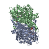
| Title | Crystal Structure of E447A Acyl-CoA Dehydrogenase FadE5 mutant from Mycobacteria smegmatis in complex with C8CoA | ||||||
 Components Components | Acyl-CoA dehydrogenase | ||||||
 Keywords Keywords | OXIDOREDUCTASE / dehydrogenase | ||||||
| Function / homology |  Function and homology information Function and homology information: / short-chain fatty acyl-CoA dehydrogenase activity / long-chain acyl-CoA dehydrogenase / long-chain fatty acyl-CoA dehydrogenase activity / medium-chain acyl-CoA dehydrogenase / short-chain acyl-CoA dehydrogenase / medium-chain fatty acyl-CoA dehydrogenase activity / fatty acid metabolic process / flavin adenine dinucleotide binding / plasma membrane Similarity search - Function | ||||||
| Biological species |  Mycobacterium smegmatis (bacteria) Mycobacterium smegmatis (bacteria) | ||||||
| Method |  X-RAY DIFFRACTION / SYNCHROTRON / FOURIER SYNTHESIS / Resolution: 2.8 Å X-RAY DIFFRACTION / SYNCHROTRON / FOURIER SYNTHESIS / Resolution: 2.8 Å | ||||||
 Authors Authors | Liu, X. / Chen, X.B. | ||||||
 Citation Citation | Journal: Proc.Natl.Acad.Sci.USA / Year: 2020 Title: Structural basis for the broad substrate specificity of two acyl-CoA dehydrogenases FadE5 from mycobacteria. Authors: Chen, X. / Chen, J. / Yan, B. / Zhang, W. / Guddat, L.W. / Liu, X. / Rao, Z. | ||||||
| History |
|
- Structure visualization
| Structure viewer | Molecule: MolmilJmol/JSmol |
|---|
- Downloads & links
-Download
| PDBx/mmCIF format | 6lq1.cif.gz | 443.5 KB | Display | PDBx/mmCIF format |
|---|---|---|---|---|
| PDB format | pdb6lq1.ent.gz | 366.4 KB | Display | PDB format |
| PDBx/mmJSON format | 6lq1.json.gz | Tree view | PDBx/mmJSON format | |
| Others |  Other downloads Other downloads |
-Validation report
| Arichive directory | https://data.pdbj.org/pub/pdb/validation_reports/lq/6lq1ftp://data.pdbj.org/pub/pdb/validation_reports/lq/6lq1 | HTTPS FTP |
|---|
-Related structure data
| Related structure data |  6kptC  6kriC  6ks9C  6ksaC  6ksbC  6kseC  6lpyC  6lq0C  6lq2C  6lq3C  6lq4C  6lq5C  6lq6C  6lq7C  6lq8C C: citing same article ( |
|---|---|
| Similar structure data |


-Links
 PDBj
PDBj


- Assembly
| Deposited unit | 
| ||||||||
|---|---|---|---|---|---|---|---|---|---|
| 1 |
| ||||||||
| Unit cell |
|
-Components
| #1: Protein | Mass: 66543.203 Da / Num. of mol.: 2 / Mutation: E447A Source method: isolated from a genetically manipulated source Source: (gene. exp.) Mycobacterium smegmatis (bacteria) / Gene: fadE5, ERS451418_00380 / Plasmid: pET28 / Production host: References: UniProt: A0A0D6G5A8, UniProt: Q3L887*PLUS, short-chain acyl-CoA dehydrogenase #2: Chemical |   Mass: 785.550 Da / Num. of mol.: 2 / Source method: obtained synthetically / Formula: C27H33N9O15P2 / Feature type: SUBJECT OF INVESTIGATION / Comment: FAD*YM Mass: 785.550 Da / Num. of mol.: 2 / Source method: obtained synthetically / Formula: C27H33N9O15P2 / Feature type: SUBJECT OF INVESTIGATION / Comment: FAD*YM#3: Chemical |   Mass: 144.211 Da / Num. of mol.: 2 Mass: 144.211 Da / Num. of mol.: 2Source method: isolated from a genetically manipulated source Formula: C8H16O2 #4: Chemical |   Mass: 767.534 Da / Num. of mol.: 2 Mass: 767.534 Da / Num. of mol.: 2Source method: isolated from a genetically manipulated source Formula: C21H36N7O16P3S #5: Water | ChemComp-HOH / |  Mass: 18.015 Da / Num. of mol.: 254 / Source method: isolated from a natural source / Formula: H2O Mass: 18.015 Da / Num. of mol.: 254 / Source method: isolated from a natural source / Formula: H2OHas ligand of interest | Y | |
|---|
-Experimental details
-Experiment
| Experiment | Method: X-RAY DIFFRACTION / Number of used crystals: 1 |
|---|
- Sample preparation
| Crystal | Density Matthews: 2.83 Å3/Da / Density % sol: 56.48 % |
|---|---|
| Crystal grow | Temperature: 298 K / Method: evaporation / pH: 7 Details: 0.1 M Hepes sodium (pH 7.0), 2% v/v PEG 400, 2 M (NH4)2SO4, 1 mM FAD and 1.2 mM C18CoA |
-Data collection
| Diffraction | Mean temperature: 100 K / Serial crystal experiment: N | ||||||||||||||||||||||||||||||||||||||||||||||||||||||||||||||||||||||||||||||||||||||||||||||||||||||||||||||||||||||||||||||||||||||||||||||||||||||||||||||||||||||||||||||||||||||||||||||||||||||||||||||||||
|---|---|---|---|---|---|---|---|---|---|---|---|---|---|---|---|---|---|---|---|---|---|---|---|---|---|---|---|---|---|---|---|---|---|---|---|---|---|---|---|---|---|---|---|---|---|---|---|---|---|---|---|---|---|---|---|---|---|---|---|---|---|---|---|---|---|---|---|---|---|---|---|---|---|---|---|---|---|---|---|---|---|---|---|---|---|---|---|---|---|---|---|---|---|---|---|---|---|---|---|---|---|---|---|---|---|---|---|---|---|---|---|---|---|---|---|---|---|---|---|---|---|---|---|---|---|---|---|---|---|---|---|---|---|---|---|---|---|---|---|---|---|---|---|---|---|---|---|---|---|---|---|---|---|---|---|---|---|---|---|---|---|---|---|---|---|---|---|---|---|---|---|---|---|---|---|---|---|---|---|---|---|---|---|---|---|---|---|---|---|---|---|---|---|---|---|---|---|---|---|---|---|---|---|---|---|---|---|---|---|---|---|
| Diffraction source | Source: SYNCHROTRON / Site: SSRF  / Beamline: BL19U1 / Wavelength: 0.9785 Å / Beamline: BL19U1 / Wavelength: 0.9785 Å | ||||||||||||||||||||||||||||||||||||||||||||||||||||||||||||||||||||||||||||||||||||||||||||||||||||||||||||||||||||||||||||||||||||||||||||||||||||||||||||||||||||||||||||||||||||||||||||||||||||||||||||||||||
| Detector | Type: DECTRIS PILATUS3 S 6M / Detector: PIXEL / Date: Nov 17, 2019 | ||||||||||||||||||||||||||||||||||||||||||||||||||||||||||||||||||||||||||||||||||||||||||||||||||||||||||||||||||||||||||||||||||||||||||||||||||||||||||||||||||||||||||||||||||||||||||||||||||||||||||||||||||
| Radiation | Protocol: SINGLE WAVELENGTH / Monochromatic (M) / Laue (L): M / Scattering type: x-ray | ||||||||||||||||||||||||||||||||||||||||||||||||||||||||||||||||||||||||||||||||||||||||||||||||||||||||||||||||||||||||||||||||||||||||||||||||||||||||||||||||||||||||||||||||||||||||||||||||||||||||||||||||||
| Radiation wavelength | Wavelength: 0.9785 Å / Relative weight: 1 | ||||||||||||||||||||||||||||||||||||||||||||||||||||||||||||||||||||||||||||||||||||||||||||||||||||||||||||||||||||||||||||||||||||||||||||||||||||||||||||||||||||||||||||||||||||||||||||||||||||||||||||||||||
| Reflection | Resolution: 2.8→19.668 Å / Num. obs: 37912 / % possible obs: 99.6 % / Redundancy: 13.02 % / Biso Wilson estimate: 18.818 Å2 / CC1/2: 0.995 / Rmerge(I) obs: 0.199 / Rrim(I) all: 0.207 / Χ2: 0.947 / Net I/σ(I): 16.27 / Num. measured all: 493628 | ||||||||||||||||||||||||||||||||||||||||||||||||||||||||||||||||||||||||||||||||||||||||||||||||||||||||||||||||||||||||||||||||||||||||||||||||||||||||||||||||||||||||||||||||||||||||||||||||||||||||||||||||||
| Reflection shell | Diffraction-ID: 1
|
- Processing
| Software |
| ||||||||||||||||||||||||||||||||||||||||||||||||||||||||||||||||||||||||||||||
|---|---|---|---|---|---|---|---|---|---|---|---|---|---|---|---|---|---|---|---|---|---|---|---|---|---|---|---|---|---|---|---|---|---|---|---|---|---|---|---|---|---|---|---|---|---|---|---|---|---|---|---|---|---|---|---|---|---|---|---|---|---|---|---|---|---|---|---|---|---|---|---|---|---|---|---|---|---|---|---|
| Refinement | Method to determine structure: FOURIER SYNTHESIS / Resolution: 2.8→19.668 Å / SU ML: 0.29 / Cross valid method: THROUGHOUT / σ(F): 1.86 / Phase error: 23.03
| ||||||||||||||||||||||||||||||||||||||||||||||||||||||||||||||||||||||||||||||
| Solvent computation | Shrinkage radii: 0.9 Å / VDW probe radii: 1.11 Å | ||||||||||||||||||||||||||||||||||||||||||||||||||||||||||||||||||||||||||||||
| Displacement parameters | Biso max: 76.79 Å2 / Biso mean: 22.2655 Å2 / Biso min: 7.6 Å2 | ||||||||||||||||||||||||||||||||||||||||||||||||||||||||||||||||||||||||||||||
| Refinement step | Cycle: final / Resolution: 2.8→19.668 Å
| ||||||||||||||||||||||||||||||||||||||||||||||||||||||||||||||||||||||||||||||
| LS refinement shell | Refine-ID: X-RAY DIFFRACTION / Rfactor Rfree error: 0
|
