 Movie
Movie Controller

[English] 日本語
 Yorodumi
- PDB-6lpw: Structure of Spermidine disinapoyl transferases(SDT) from Arabido... -
+ Open data

- Basic information
| Entry | Database: PDB / ID: 6lpw | ||||||
|---|---|---|---|---|---|---|---|
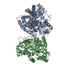
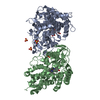
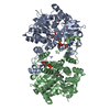
| Title | Structure of Spermidine disinapoyl transferases(SDT) from Arabidopsis thaliana | ||||||
 Components Components | Spermidine sinapoyl-CoA acyltransferase | ||||||
 Keywords Keywords | PLANT PROTEIN / BAHD transferase / phenolamides / spermidine / putrescine / multisite-acylation / molecular mechanism / sequence similarity network | ||||||
| Function / homology |  Function and homology information Function and homology informationspermidine disinapoyl transferase / sinapoyl spermidine:sinapoyl CoA N-acyltransferase activity / spermidine:sinapoyl CoA N-acyltransferase activity / polyamine biosynthetic process / spermidine metabolic process Similarity search - Function | ||||||
| Biological species |  | ||||||
| Method |  X-RAY DIFFRACTION / SYNCHROTRON / MOLECULAR REPLACEMENT / Resolution: 2.401 Å X-RAY DIFFRACTION / SYNCHROTRON / MOLECULAR REPLACEMENT / Resolution: 2.401 Å | ||||||
 Authors Authors | Wang, C.Y. / Zhang, P. | ||||||
 Citation Citation | Journal: Front Plant Sci / Year: 2020 Title: Structural and Biochemical Insights Into Two BAHD Acyltransferases ( At SHT and At SDT) Involved in Phenolamide Biosynthesis. Authors: Wang, C. / Li, J. / Ma, M. / Lin, Z. / Hu, W. / Lin, W. / Zhang, P. | ||||||
| History |
|
- Structure visualization
| Structure viewer | Molecule: MolmilJmol/JSmol |
|---|
- Downloads & links
-Download
| PDBx/mmCIF format | 6lpw.cif.gz | 181.8 KB | Display | PDBx/mmCIF format |
|---|---|---|---|---|
| PDB format | pdb6lpw.ent.gz | 144.6 KB | Display | PDB format |
| PDBx/mmJSON format | 6lpw.json.gz | Tree view | PDBx/mmJSON format | |
| Others |  Other downloads Other downloads |
-Validation report
| Arichive directory | https://data.pdbj.org/pub/pdb/validation_reports/lp/6lpwftp://data.pdbj.org/pub/pdb/validation_reports/lp/6lpw | HTTPS FTP |
|---|
-Related structure data
| Related structure data | 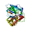 6lpvC  4g0bS C: citing same article ( S: Starting model for refinement |
|---|---|
| Similar structure data |


-Links
 PDBj
PDBj


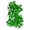
- Assembly
| Deposited unit | 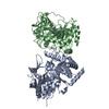
| ||||||||
|---|---|---|---|---|---|---|---|---|---|
| 1 | 
| ||||||||
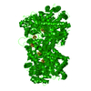
| Unit cell |
|
-Components
| #1: Protein | Mass: 50432.699 Da / Num. of mol.: 2 Source method: isolated from a genetically manipulated source Source: (gene. exp.)  References: UniProt: O80467, spermidine disinapoyl transferase #2: Chemical | ChemComp-SPD / |   Mass: 145.246 Da / Num. of mol.: 1 / Source method: obtained synthetically / Formula: C7H19N3 / Feature type: SUBJECT OF INVESTIGATION Mass: 145.246 Da / Num. of mol.: 1 / Source method: obtained synthetically / Formula: C7H19N3 / Feature type: SUBJECT OF INVESTIGATION#3: Water | ChemComp-HOH / |  Mass: 18.015 Da / Num. of mol.: 124 / Source method: isolated from a natural source / Formula: H2O Mass: 18.015 Da / Num. of mol.: 124 / Source method: isolated from a natural source / Formula: H2OHas ligand of interest | Y | |
|---|
-Experimental details
-Experiment
| Experiment | Method: X-RAY DIFFRACTION / Number of used crystals: 1 |
|---|
- Sample preparation
| Crystal | Density Matthews: 2.11 Å3/Da / Density % sol: 41.7 % |
|---|---|
| Crystal grow | Temperature: 293.15 K / Method: vapor diffusion, sitting drop / Details: 0.1 M MES (pH 6.5), 25% W/V PEG 4000 at 20 C |
-Data collection
| Diffraction | Mean temperature: 77 K / Serial crystal experiment: N |
|---|---|
| Diffraction source | Source: SYNCHROTRON / Site: APS  / Beamline: 17-BM / Wavelength: 0.987 Å / Beamline: 17-BM / Wavelength: 0.987 Å |
| Detector | Type: DECTRIS EIGER X 500K / Detector: PIXEL / Date: Jan 9, 2013 |
| Radiation | Protocol: SINGLE WAVELENGTH / Monochromatic (M) / Laue (L): M / Scattering type: x-ray |
| Radiation wavelength | Wavelength: 0.987 Å / Relative weight: 1 |
| Reflection | Resolution: 2.4→50 Å / Num. obs: 32701 / % possible obs: 95.82 % / Redundancy: 3 % / CC1/2: 0.09 / Net I/σ(I): 15 |
| Reflection shell | Resolution: 2.49→2.49 Å / CC1/2: 0.09 |
- Processing
| Software |
| ||||||||||||||||||||||||||||||||||||||||||||||||||||||||||||||||||||||||||||||
|---|---|---|---|---|---|---|---|---|---|---|---|---|---|---|---|---|---|---|---|---|---|---|---|---|---|---|---|---|---|---|---|---|---|---|---|---|---|---|---|---|---|---|---|---|---|---|---|---|---|---|---|---|---|---|---|---|---|---|---|---|---|---|---|---|---|---|---|---|---|---|---|---|---|---|---|---|---|---|---|
| Refinement | Method to determine structure: MOLECULAR REPLACEMENT Starting model: 4G0B Resolution: 2.401→48.161 Å / SU ML: 0.27 / Cross valid method: THROUGHOUT / σ(F): 1.35 / Phase error: 24.98
| ||||||||||||||||||||||||||||||||||||||||||||||||||||||||||||||||||||||||||||||
| Solvent computation | Shrinkage radii: 0.9 Å / VDW probe radii: 1.11 Å | ||||||||||||||||||||||||||||||||||||||||||||||||||||||||||||||||||||||||||||||
| Displacement parameters | Biso max: 140.22 Å2 / Biso mean: 41.4353 Å2 / Biso min: 15.77 Å2 | ||||||||||||||||||||||||||||||||||||||||||||||||||||||||||||||||||||||||||||||
| Refinement step | Cycle: final / Resolution: 2.401→48.161 Å
| ||||||||||||||||||||||||||||||||||||||||||||||||||||||||||||||||||||||||||||||
| LS refinement shell | Refine-ID: X-RAY DIFFRACTION / Rfactor Rfree error: 0
|
