 Movie
Movie Controller

[English] 日本語
 Yorodumi
- PDB-2i3o: Crystal structure of gamma-glutamyl transferase related protein f... -
+ Open data

- Basic information
| Entry | Database: PDB / ID: 2i3o | ||||||
|---|---|---|---|---|---|---|---|
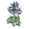
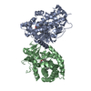
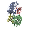
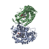
| Title | Crystal structure of gamma-glutamyl transferase related protein from Thermoplasma acidophilum | ||||||
 Components Components | Gamma-glutamyltransferase related protein | ||||||
 Keywords Keywords | TRANSFERASE / Gamma-glutamyl transferase related protein / Thermoplasma acidophilum / 6324d / Structural Genomics / PSI / Protein Structure Initiative / New York SGX Research Center for Structural Genomics / NYSGXRC | ||||||
| Function / homology | : / Gamma-glutamyltranspeptidase / Gamma-glutamyltranspeptidase, small subunit / Nucleophile aminohydrolases, N-terminal / Gamma-glutamyltransferase related protein Function and homology information Function and homology information | ||||||
| Biological species |   Thermoplasma acidophilum (acidophilic) Thermoplasma acidophilum (acidophilic) | ||||||
| Method |  X-RAY DIFFRACTION / SYNCHROTRON / SAD / Resolution: 2.03 Å X-RAY DIFFRACTION / SYNCHROTRON / SAD / Resolution: 2.03 Å | ||||||
 Authors Authors | Rao, K.N. / Eswaramoorthy, S. / Burley, S.K. / Swaminathan, S. / New York SGX Research Center for Structural Genomics (NYSGXRC) | ||||||
 Citation Citation | Journal: To be Published Title: Crystal structure of gamma-glutamyl transferase related protein from Thermoplasma acidophilum Authors: Rao, K.N. / Eswaramoorthy, S. / Burley, S.K. / Swaminathan, S. | ||||||
| History |
|
- Structure visualization
| Structure viewer | Molecule: MolmilJmol/JSmol |
|---|
- Downloads & links
-Download
| PDBx/mmCIF format | 2i3o.cif.gz | 403.7 KB | Display | PDBx/mmCIF format |
|---|---|---|---|---|
| PDB format | pdb2i3o.ent.gz | 331.3 KB | Display | PDB format |
| PDBx/mmJSON format | 2i3o.json.gz | Tree view | PDBx/mmJSON format | |
| Others |  Other downloads Other downloads |
-Validation report
| Arichive directory | https://data.pdbj.org/pub/pdb/validation_reports/i3/2i3oftp://data.pdbj.org/pub/pdb/validation_reports/i3/2i3o | HTTPS FTP |
|---|
-Related structure data
| Similar structure data | |
|---|---|
| Other databases |


-Links
 PDBj
PDBj


- Assembly
| Deposited unit | 
| ||||||||
|---|---|---|---|---|---|---|---|---|---|
| 1 | 
| ||||||||
| 2 | 
| ||||||||
| Unit cell |
|
-Components
| #1: Protein | Mass: 57578.977 Da / Num. of mol.: 4 Source method: isolated from a genetically manipulated source Source: (gene. exp.) Thermoplasma acidophilum (acidophilic) / Production host:  #2: Water | ChemComp-HOH / |  Mass: 18.015 Da / Num. of mol.: 887 / Source method: isolated from a natural source / Formula: H2O Mass: 18.015 Da / Num. of mol.: 887 / Source method: isolated from a natural source / Formula: H2OHas protein modification | Y | |
|---|
-Experimental details
-Experiment
| Experiment | Method: X-RAY DIFFRACTION / Number of used crystals: 1 |
|---|
- Sample preparation
| Crystal | Density Matthews: 2.69 Å3/Da / Density % sol: 54.34 % |
|---|---|
| Crystal grow | Temperature: 293 K / Method: vapor diffusion, sitting drop / pH: 5.5 Details: Magnesium Formate, pH 5.5, VAPOR DIFFUSION, SITTING DROP, temperature 293K |
-Data collection
| Diffraction | Mean temperature: 100 K |
|---|---|
| Diffraction source | Source: SYNCHROTRON / Site: NSLS  / Beamline: X12C / Wavelength: 0.9795 Å / Beamline: X12C / Wavelength: 0.9795 Å |
| Detector | Type: ADSC QUANTUM 210 / Detector: CCD / Date: Jun 14, 2006 / Details: Mirrors |
| Radiation | Monochromator: Si(111) / Protocol: SINGLE WAVELENGTH / Monochromatic (M) / Laue (L): M / Scattering type: x-ray |
| Radiation wavelength | Wavelength: 0.9795 Å / Relative weight: 1 |
| Reflection | Resolution: 2.03→50 Å / Num. all: 156657 / Num. obs: 156657 / % possible obs: 99.6 % / Observed criterion σ(F): 0 / Observed criterion σ(I): 0 / Redundancy: 7.4 % / Biso Wilson estimate: 11.4 Å2 / Rmerge(I) obs: 0.095 / Net I/σ(I): 11.5 |
| Reflection shell | Resolution: 2.03→2.1 Å / Redundancy: 6.5 % / Mean I/σ(I) obs: 3 / Num. unique all: 15286 / Rsym value: 0.38 / % possible all: 97.9 |
- Processing
| Software |
| ||||||||||||||||||||||||||||||||||||
|---|---|---|---|---|---|---|---|---|---|---|---|---|---|---|---|---|---|---|---|---|---|---|---|---|---|---|---|---|---|---|---|---|---|---|---|---|---|
| Refinement | Method to determine structure: SAD / Resolution: 2.03→30.47 Å / Rfactor Rfree error: 0.003 / Data cutoff high absF: 92146.53 / Data cutoff low absF: 0 / Isotropic thermal model: RESTRAINED / Cross valid method: THROUGHOUT / σ(F): 0 / Stereochemistry target values: Engh & Huber Details: Residues listed in remark 465 and atoms listed in remark 470 were not modeled due to lack of electron density. Residue ASN254 in all four chains does not have good geometry. However, they ...Details: Residues listed in remark 465 and atoms listed in remark 470 were not modeled due to lack of electron density. Residue ASN254 in all four chains does not have good geometry. However, they fit the electron density very well. Residual densities near the active sites have been modeled as water (39, 40, 45, 167) but they could be metal ions. N terminal Selenomethionine is visible only in chains A and D.
| ||||||||||||||||||||||||||||||||||||
| Solvent computation | Solvent model: FLAT MODEL / Bsol: 40.6224 Å2 / ksol: 0.369856 e/Å3 | ||||||||||||||||||||||||||||||||||||
| Displacement parameters | Biso mean: 19.7 Å2
| ||||||||||||||||||||||||||||||||||||
| Refine analyze |
| ||||||||||||||||||||||||||||||||||||
| Refinement step | Cycle: LAST / Resolution: 2.03→30.47 Å
| ||||||||||||||||||||||||||||||||||||
| Refine LS restraints |
| ||||||||||||||||||||||||||||||||||||
| LS refinement shell | Resolution: 2.03→2.16 Å / Rfactor Rfree error: 0.01 / Total num. of bins used: 6
|
