 Movie
Movie Controller

[English] 日本語
 Yorodumi
- PDB-6lgi: Bombyx mori GH13 sucrose hydrolase mutant E322Q covalent intermed... -
+ Open data

- Basic information
| Entry | Database: PDB / ID: 6lgi | ||||||
|---|---|---|---|---|---|---|---|
| Title | Bombyx mori GH13 sucrose hydrolase mutant E322Q covalent intermediate complexed with fructose | ||||||
 Components Components | Sucrose hydrolase | ||||||
 Keywords Keywords | HYDROLASE / Sucrose / Glycoside hydrolase / GH13 | ||||||
| Function / homology |  Function and homology information Function and homology informationalpha-glucosidase / alpha-1,4-glucosidase activity / hydrolase activity, acting on glycosyl bonds / carbohydrate metabolic process / metal ion binding Similarity search - Function | ||||||
| Biological species |  | ||||||
| Method |  X-RAY DIFFRACTION / SYNCHROTRON / MOLECULAR REPLACEMENT / Resolution: 1.6 Å X-RAY DIFFRACTION / SYNCHROTRON / MOLECULAR REPLACEMENT / Resolution: 1.6 Å | ||||||
 Authors Authors | Miyazaki, T. | ||||||
| Funding support |  Japan, 1items Japan, 1items
| ||||||
 Citation Citation | Journal: J.Biol.Chem. / Year: 2020 Title: Structure-function analysis of silkworm sucrose hydrolase uncovers the mechanism of substrate specificity in GH13 subfamily 17exo-alpha-glucosidases. Authors: Miyazaki, T. / Park, E.Y. | ||||||
| History |
|
- Structure visualization
| Structure viewer | Molecule: MolmilJmol/JSmol |
|---|
- Downloads & links
-Download
| PDBx/mmCIF format | 6lgi.cif.gz | 272.3 KB | Display | PDBx/mmCIF format |
|---|---|---|---|---|
| PDB format | pdb6lgi.ent.gz | 213.4 KB | Display | PDB format |
| PDBx/mmJSON format | 6lgi.json.gz | Tree view | PDBx/mmJSON format | |
| Others |  Other downloads Other downloads |
-Validation report
| Arichive directory | https://data.pdbj.org/pub/pdb/validation_reports/lg/6lgiftp://data.pdbj.org/pub/pdb/validation_reports/lg/6lgi | HTTPS FTP |
|---|
-Related structure data
| Related structure data |  6lgaC  6lgbC  6lgcC  6lgdC  6lgeC 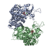 6lgfC 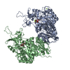 6lggC 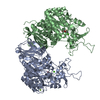 6lghC 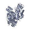 5brqS S: Starting model for refinement C: citing same article ( |
|---|---|
| Similar structure data |


-Links
 PDBj
PDBj


- Assembly
| Deposited unit | 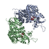
| ||||||||
|---|---|---|---|---|---|---|---|---|---|
| 1 |
| ||||||||
| Unit cell |
|
-Components
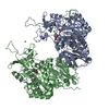
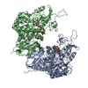
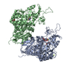
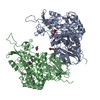
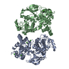
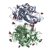
-Protein , 1 types, 2 molecules AB
| #1: Protein | Mass: 68738.219 Da / Num. of mol.: 2 / Mutation: E322Q Source method: isolated from a genetically manipulated source Source: (gene. exp.)  |
|---|
-Sugars , 3 types, 6 molecules 
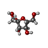

| #4: Sugar |  Type: D-saccharide, beta linking / Mass: 180.156 Da / Num. of mol.: 2 Type: D-saccharide, beta linking / Mass: 180.156 Da / Num. of mol.: 2Source method: isolated from a genetically manipulated source Formula: C6H12O6 / Feature type: SUBJECT OF INVESTIGATION #5: Sugar |  Type: D-saccharide, beta linking / Mass: 180.156 Da / Num. of mol.: 2 Type: D-saccharide, beta linking / Mass: 180.156 Da / Num. of mol.: 2Source method: isolated from a genetically manipulated source Formula: C6H12O6 / Feature type: SUBJECT OF INVESTIGATION #6: Sugar |  Type: D-saccharide, beta linking / Mass: 180.156 Da / Num. of mol.: 2 Type: D-saccharide, beta linking / Mass: 180.156 Da / Num. of mol.: 2Source method: isolated from a genetically manipulated source Formula: C6H12O6 / Feature type: SUBJECT OF INVESTIGATION |
|---|
-Non-polymers , 4 types, 992 molecules 


| #2: Chemical | ChemComp-MG /  Mass: 24.305 Da / Num. of mol.: 5 / Source method: obtained synthetically / Formula: Mg / Feature type: SUBJECT OF INVESTIGATION Mass: 24.305 Da / Num. of mol.: 5 / Source method: obtained synthetically / Formula: Mg / Feature type: SUBJECT OF INVESTIGATION#3: Chemical |  Mass: 40.078 Da / Num. of mol.: 2 / Source method: obtained synthetically / Formula: Ca / Feature type: SUBJECT OF INVESTIGATION Mass: 40.078 Da / Num. of mol.: 2 / Source method: obtained synthetically / Formula: Ca / Feature type: SUBJECT OF INVESTIGATION#7: Chemical |  Mass: 59.044 Da / Num. of mol.: 2 / Source method: obtained synthetically / Formula: C2H3O2 Mass: 59.044 Da / Num. of mol.: 2 / Source method: obtained synthetically / Formula: C2H3O2#8: Water | ChemComp-HOH / | Mass: 18.015 Da / Num. of mol.: 983 / Source method: isolated from a natural source / Formula: H2O |
|---|
-Details
| Has ligand of interest | Y |
|---|---|
| Has protein modification | Y |
-Experimental details
-Experiment
| Experiment | Method: X-RAY DIFFRACTION / Number of used crystals: 1 |
|---|
- Sample preparation
| Crystal | Density Matthews: 2.69 Å3/Da / Density % sol: 54.2 % |
|---|---|
| Crystal grow | Temperature: 293 K / Method: vapor diffusion, hanging drop Details: 10% PEG 3350, 0.2 M magnesium acetate, 10 mM alpha-glucopyranosyl fluoride, 10 mM fructose |
-Data collection
| Diffraction | Mean temperature: 100 K / Serial crystal experiment: N |
|---|---|
| Diffraction source | Source: SYNCHROTRON / Site: Photon Factory / Beamline: AR-NW12A / Wavelength: 1 Å |
| Detector | Type: DECTRIS PILATUS3 S 2M / Detector: PIXEL / Date: Jun 23, 2019 |
| Radiation | Protocol: SINGLE WAVELENGTH / Monochromatic (M) / Laue (L): M / Scattering type: x-ray |
| Radiation wavelength | Wavelength: 1 Å / Relative weight: 1 |
| Reflection | Resolution: 1.6→50 Å / Num. obs: 194405 / % possible obs: 99.7 % / Redundancy: 6.4 % / CC1/2: 0.999 / Rmerge(I) obs: 0.061 / Net I/σ(I): 16.9 |
| Reflection shell | Resolution: 1.6→1.69 Å / Redundancy: 6.6 % / Rmerge(I) obs: 0.65 / Mean I/σ(I) obs: 2.6 / Num. unique obs: 28198 / CC1/2: 0.833 / % possible all: 100 |
- Processing
| Software |
| ||||||||||||||||||||||||||||||||||||||||||||||||||||||||||||
|---|---|---|---|---|---|---|---|---|---|---|---|---|---|---|---|---|---|---|---|---|---|---|---|---|---|---|---|---|---|---|---|---|---|---|---|---|---|---|---|---|---|---|---|---|---|---|---|---|---|---|---|---|---|---|---|---|---|---|---|---|---|
| Refinement | Method to determine structure: MOLECULAR REPLACEMENT Starting model: 5BRQ Resolution: 1.6→42.04 Å / Cor.coef. Fo:Fc: 0.966 / Cor.coef. Fo:Fc free: 0.955 / SU B: 1.925 / SU ML: 0.065 / Cross valid method: THROUGHOUT / σ(F): 0 / ESU R: 0.079 / ESU R Free: 0.08 Details: HYDROGENS HAVE BEEN ADDED IN THE RIDING POSITIONS U VALUES : REFINED INDIVIDUALLY
| ||||||||||||||||||||||||||||||||||||||||||||||||||||||||||||
| Solvent computation | Ion probe radii: 0.8 Å / Shrinkage radii: 0.8 Å / VDW probe radii: 1.2 Å | ||||||||||||||||||||||||||||||||||||||||||||||||||||||||||||
| Displacement parameters | Biso max: 108.97 Å2 / Biso mean: 29.143 Å2 / Biso min: 17.15 Å2
| ||||||||||||||||||||||||||||||||||||||||||||||||||||||||||||
| Refinement step | Cycle: final / Resolution: 1.6→42.04 Å
| ||||||||||||||||||||||||||||||||||||||||||||||||||||||||||||
| Refine LS restraints |
| ||||||||||||||||||||||||||||||||||||||||||||||||||||||||||||
| LS refinement shell | Resolution: 1.6→1.642 Å / Rfactor Rfree error: 0 / Total num. of bins used: 20
|
