 Movie
Movie Controller


+ Open data

- Basic information
| Entry | Database: PDB / ID: 6lg2 | ||||||
|---|---|---|---|---|---|---|---|
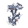
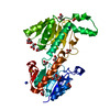
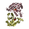
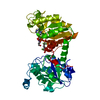
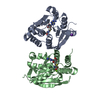
| Title | VanR bound to Vanillate | ||||||
 Components Components | Predicted transcriptional regulators | ||||||
 Keywords Keywords | TRANSCRIPTION / VanR transcription factor Vanillate | ||||||
| Function / homology | Transcription regulator PadR, N-terminal / Transcriptional regulator PadR-like family / Winged helix DNA-binding domain superfamily / Winged helix-like DNA-binding domain superfamily / 4-HYDROXY-3-METHOXYBENZOATE / Predicted transcriptional regulators Function and homology information Function and homology information | ||||||
| Biological species |  Corynebacterium glutamicum (bacteria) Corynebacterium glutamicum (bacteria) | ||||||
| Method |  X-RAY DIFFRACTION / SYNCHROTRON / MOLECULAR REPLACEMENT / Resolution: 1.6 Å X-RAY DIFFRACTION / SYNCHROTRON / MOLECULAR REPLACEMENT / Resolution: 1.6 Å | ||||||
 Authors Authors | He, Y. / Bharath, S.R. / Song, H. | ||||||
 Citation Citation | Journal: Nat Commun / Year: 2020 Title: Developing a highly efficient hydroxytyrosol whole-cell catalyst by de-bottlenecking rate-limiting steps. Authors: Yao, J. / He, Y. / Su, N. / Bharath, S.R. / Tao, Y. / Jin, J.M. / Chen, W. / Song, H. / Tang, S.Y. | ||||||
| History |
|
- Structure visualization
| Structure viewer | Molecule: MolmilJmol/JSmol |
|---|
- Downloads & links
-Download
| PDBx/mmCIF format | 6lg2.cif.gz | 100.4 KB | Display | PDBx/mmCIF format |
|---|---|---|---|---|
| PDB format | pdb6lg2.ent.gz | 69.8 KB | Display | PDB format |
| PDBx/mmJSON format | 6lg2.json.gz | Tree view | PDBx/mmJSON format | |
| Others |  Other downloads Other downloads |
-Validation report
| Arichive directory | https://data.pdbj.org/pub/pdb/validation_reports/lg/6lg2ftp://data.pdbj.org/pub/pdb/validation_reports/lg/6lg2 | HTTPS FTP |
|---|
-Related structure data
| Related structure data |  3l9fS S: Starting model for refinement |
|---|---|
| Similar structure data |


-Links
 PDBj
PDBj- Assembly
| Deposited unit | 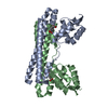
| ||||||||||||
|---|---|---|---|---|---|---|---|---|---|---|---|---|---|
| 1 |
| ||||||||||||
| Unit cell |
|
-Components
| #1: Protein | Mass: 21592.531 Da / Num. of mol.: 2 Source method: isolated from a genetically manipulated source Details: vanR transcriptional regulator Source: (gene. exp.) Corynebacterium glutamicum (strain ATCC 13032 / DSM 20300 / JCM 1318 / LMG 3730 / NCIMB 10025) (bacteria)Strain: ATCC 13032 / DSM 20300 / JCM 1318 / LMG 3730 / NCIMB 10025 Gene: Cgl2382 / Production host: #2: Chemical |   Mass: 167.139 Da / Num. of mol.: 2 / Source method: obtained synthetically / Formula: C8H7O4 / Feature type: SUBJECT OF INVESTIGATION Mass: 167.139 Da / Num. of mol.: 2 / Source method: obtained synthetically / Formula: C8H7O4 / Feature type: SUBJECT OF INVESTIGATION#3: Water | ChemComp-HOH / |  Mass: 18.015 Da / Num. of mol.: 335 / Source method: isolated from a natural source / Formula: H2O Mass: 18.015 Da / Num. of mol.: 335 / Source method: isolated from a natural source / Formula: H2OHas ligand of interest | Y | |
|---|
-Experimental details
-Experiment
| Experiment | Method: X-RAY DIFFRACTION / Number of used crystals: 1 |
|---|
- Sample preparation
| Crystal | Density Matthews: 2.14 Å3/Da / Density % sol: 42.51 % |
|---|---|
| Crystal grow | Temperature: 293 K / Method: vapor diffusion, hanging drop Details: 0.2 M Magnesium chloride 0.1 M Tris pH 8.0 12% PEG4000 |
-Data collection
| Diffraction | Mean temperature: 100 K / Serial crystal experiment: N |
|---|---|
| Diffraction source | Source: SYNCHROTRON / Site: SSRF  / Beamline: BL18U1 / Wavelength: 0.97853 Å / Beamline: BL18U1 / Wavelength: 0.97853 Å |
| Detector | Type: DECTRIS PILATUS 6M / Detector: PIXEL / Date: May 13, 2016 |
| Radiation | Protocol: SINGLE WAVELENGTH / Monochromatic (M) / Laue (L): M / Scattering type: x-ray |
| Radiation wavelength | Wavelength: 0.97853 Å / Relative weight: 1 |
| Reflection | Resolution: 1.6→29.57 Å / Num. obs: 44461 / % possible obs: 93 % / Redundancy: 1.8 % / Biso Wilson estimate: 15.22 Å2 / CC1/2: 0.987 / Rmerge(I) obs: 0.097 / Net I/σ(I): 6.3 |
| Reflection shell | Resolution: 1.6→1.63 Å / Rmerge(I) obs: 0.69 / Mean I/σ(I) obs: 1.2 / Num. unique obs: 2195 / CC1/2: 0.434 |
- Processing
| Software |
| |||||||||||||||||||||||||||||||||||||||||||||||||||||||||||||||||||||||||||||||||||||||||||||||||||||||||||||||||||||||
|---|---|---|---|---|---|---|---|---|---|---|---|---|---|---|---|---|---|---|---|---|---|---|---|---|---|---|---|---|---|---|---|---|---|---|---|---|---|---|---|---|---|---|---|---|---|---|---|---|---|---|---|---|---|---|---|---|---|---|---|---|---|---|---|---|---|---|---|---|---|---|---|---|---|---|---|---|---|---|---|---|---|---|---|---|---|---|---|---|---|---|---|---|---|---|---|---|---|---|---|---|---|---|---|---|---|---|---|---|---|---|---|---|---|---|---|---|---|---|---|---|
| Refinement | Method to determine structure: MOLECULAR REPLACEMENT Starting model: 3L9F Resolution: 1.6→29.57 Å / SU ML: 0.1921 / Cross valid method: FREE R-VALUE / σ(F): 1.35 / Phase error: 24.0677
| |||||||||||||||||||||||||||||||||||||||||||||||||||||||||||||||||||||||||||||||||||||||||||||||||||||||||||||||||||||||
| Solvent computation | Shrinkage radii: 0.9 Å / VDW probe radii: 1.11 Å | |||||||||||||||||||||||||||||||||||||||||||||||||||||||||||||||||||||||||||||||||||||||||||||||||||||||||||||||||||||||
| Displacement parameters | Biso mean: 18.32 Å2 | |||||||||||||||||||||||||||||||||||||||||||||||||||||||||||||||||||||||||||||||||||||||||||||||||||||||||||||||||||||||
| Refinement step | Cycle: LAST / Resolution: 1.6→29.57 Å
| |||||||||||||||||||||||||||||||||||||||||||||||||||||||||||||||||||||||||||||||||||||||||||||||||||||||||||||||||||||||
| Refine LS restraints |
| |||||||||||||||||||||||||||||||||||||||||||||||||||||||||||||||||||||||||||||||||||||||||||||||||||||||||||||||||||||||
| LS refinement shell |
|
