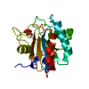
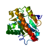
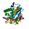
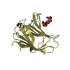
 Movie
Movie Controller


+ Open data

- Basic information
| Entry | Database: PDB / ID: 6lfu | |||||||||
|---|---|---|---|---|---|---|---|---|---|---|
| Title | Poa1p F152A mutant in complex with ADP-ribose | |||||||||
 Components Components | ADP-ribose 1''-phosphate phosphatase | |||||||||
 Keywords Keywords | HYDROLASE / deacetylase / macro domain | |||||||||
| Function / homology |  Function and homology information Function and homology information: / purine nucleoside metabolic process / tRNA splicing, via endonucleolytic cleavage and ligation / ADP-ribose 1''-phosphate phosphatase / Hydrolases; Glycosylases; Hydrolysing N-glycosyl compounds / phosphatase activity / DNA damage response / nucleoplasm Similarity search - Function | |||||||||
| Biological species |  | |||||||||
| Method |  X-RAY DIFFRACTION / SYNCHROTRON / MOLECULAR REPLACEMENT / Resolution: 3.123 Å X-RAY DIFFRACTION / SYNCHROTRON / MOLECULAR REPLACEMENT / Resolution: 3.123 Å | |||||||||
 Authors Authors | Chiu, Y.C. / Hsu, C.H. | |||||||||
| Funding support |  Taiwan, 2items Taiwan, 2items
| |||||||||
 Citation Citation | Journal: Acs Catalysis / Year: 2021 Title: Expanding the Substrate Specificity of Macro Domains toward 3''-Isomer of O-Acetyl-ADP-ribose Authors: Chiu, Y.C. / Tseng, M.C. / Hsu, C.H. | |||||||||
| History |
|
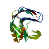
- Structure visualization
| Structure viewer | Molecule: MolmilJmol/JSmol |
|---|
- Downloads & links
-Download
| PDBx/mmCIF format | 6lfu.cif.gz | 80.8 KB | Display | PDBx/mmCIF format |
|---|---|---|---|---|
| PDB format | pdb6lfu.ent.gz | 58.2 KB | Display | PDB format |
| PDBx/mmJSON format | 6lfu.json.gz | Tree view | PDBx/mmJSON format | |
| Others |  Other downloads Other downloads |
-Validation report
| Arichive directory | https://data.pdbj.org/pub/pdb/validation_reports/lf/6lfuftp://data.pdbj.org/pub/pdb/validation_reports/lf/6lfu | HTTPS FTP |
|---|
-Related structure data
| Related structure data |  6lfqSC 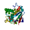 6lfrC  6lfsC 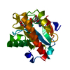 6lftC S: Starting model for refinement C: citing same article ( |
|---|---|
| Similar structure data |


-Links
 PDBj
PDBj
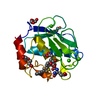
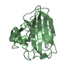
- Assembly
| Deposited unit | 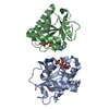
| ||||||||
|---|---|---|---|---|---|---|---|---|---|
| 1 | 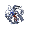
| ||||||||
| 2 | 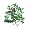
| ||||||||
| Unit cell |
|
-Components
| #1: Protein | Mass: 20167.994 Da / Num. of mol.: 2 / Mutation: F152A Source method: isolated from a genetically manipulated source Source: (gene. exp.)  References: UniProt: P38218, ADP-ribose 1''-phosphate phosphatase, Hydrolases; Glycosylases; Hydrolysing N-glycosyl compounds #2: Chemical |   Mass: 559.316 Da / Num. of mol.: 2 / Source method: obtained synthetically / Formula: C15H23N5O14P2 / Feature type: SUBJECT OF INVESTIGATION Mass: 559.316 Da / Num. of mol.: 2 / Source method: obtained synthetically / Formula: C15H23N5O14P2 / Feature type: SUBJECT OF INVESTIGATIONHas ligand of interest | Y | |
|---|
-Experimental details
-Experiment
| Experiment | Method: X-RAY DIFFRACTION / Number of used crystals: 1 |
|---|
- Sample preparation
| Crystal | Density Matthews: 2.2 Å3/Da / Density % sol: 44.11 % |
|---|---|
| Crystal grow | Temperature: 283 K / Method: vapor diffusion, sitting drop / Details: Citric acid, PEG 3350, Glycerol |
-Data collection
| Diffraction | Mean temperature: 100 K / Serial crystal experiment: N |
|---|---|
| Diffraction source | Source: SYNCHROTRON / Site: NSRRC / Beamline: BL13B1 / Wavelength: 1 Å |
| Detector | Type: RAYONIX MX300-HS / Detector: CCD / Date: Nov 2, 2018 |
| Radiation | Protocol: SINGLE WAVELENGTH / Monochromatic (M) / Laue (L): M / Scattering type: x-ray |
| Radiation wavelength | Wavelength: 1 Å / Relative weight: 1 |
| Reflection | Resolution: 3.123→24.884 Å / Num. obs: 5850 / % possible obs: 98 % / Redundancy: 3.5 % / CC1/2: 0.985 / Net I/σ(I): 20.5 |
| Reflection shell | Resolution: 3.123→3.4361 Å / CC1/2: 0.953 |
- Processing
| Software |
| ||||||||||||||||||||||||||||||
|---|---|---|---|---|---|---|---|---|---|---|---|---|---|---|---|---|---|---|---|---|---|---|---|---|---|---|---|---|---|---|---|
| Refinement | Method to determine structure: MOLECULAR REPLACEMENT Starting model: 6LFQ Resolution: 3.123→24.884 Å / SU ML: 0.44 / Cross valid method: THROUGHOUT / σ(F): 1.36 / Phase error: 27.69
| ||||||||||||||||||||||||||||||
| Solvent computation | Shrinkage radii: 0.9 Å / VDW probe radii: 1.11 Å | ||||||||||||||||||||||||||||||
| Displacement parameters | Biso max: 110.92 Å2 / Biso mean: 50.6133 Å2 / Biso min: 19.33 Å2 | ||||||||||||||||||||||||||||||
| Refinement step | Cycle: final / Resolution: 3.123→24.884 Å
| ||||||||||||||||||||||||||||||
| LS refinement shell | Refine-ID: X-RAY DIFFRACTION / Rfactor Rfree error: 0
|
