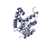
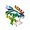
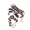
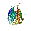
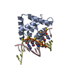
A: DNA-binding protein SATB1 B: DNA-binding protein SATB1 C: DNA (5'-D(*GP*(C7R)P*(PST)P*AP*AP*TP*AP*TP*AP*TP*GP*C)-3') D: DNA (5'-D(*GP*CP*AP*TP*(AS)P*(PST)P*(AS)P*(PST)P*TP*AP*GP*C)-3')
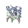
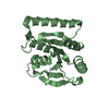
A: DNA-binding protein SATB1 C: DNA (5'-D(*GP*(C7R)P*(PST)P*AP*AP*TP*AP*TP*AP*TP*GP*C)-3') D: DNA (5'-D(*GP*CP*AP*TP*(AS)P*(PST)P*(AS)P*(PST)P*TP*AP*GP*C)-3')
defined by author&software
Evidence: isothermal titration calorimetry, 1:1 binding stoichiometry for the protein and the double-stranded DNA was indicated in solution.
Resolution: 1.79→19.63 Å / Cor.coef. Fo:Fc: 0.925 / Cor.coef. Fo:Fc free: 0.882 / SU B: 3.538 / SU ML: 0.11 / Cross valid method: THROUGHOUT / ESU R: 0.181 / ESU R Free: 0.163 / Stereochemistry target values: MAXIMUM LIKELIHOOD / Details: HYDROGENS HAVE BEEN ADDED IN THE RIDING POSITIONS
Rfactor
Num. reflection
% reflection
Selection details
Rfree
0.27122
1112
5.6 %
RANDOM
Rwork
0.22962
-
-
-
obs
0.23207
18596
93.29 %
-
Solvent computation
Ion probe radii: 0.8 Å / Shrinkage radii: 0.8 Å / VDW probe radii: 1.2 Å / Solvent model: MASK
 Movie
Movie Controller

 Yorodumi
 Open data
 Basic information
 Components
 Keywords
 Function and homology information
 Homo sapiens (human)
 X-RAY DIFFRACTION /
  Authors
 Japan, 1items
  Citation
 Structure visualization
 Downloads & links
 Other downloads
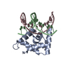


 PDBj
PDBj


 Assembly
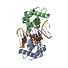

 Mass: 18.015 Da / Num. of mol.: 69 / Source method: isolated from a natural source / Formula: H2O
 Sample preparation
 Processing