 Movie
Movie Controller


+ Open data

- Basic information
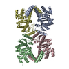
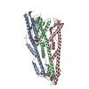
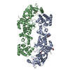
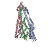
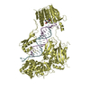
| Entry | Database: PDB / ID: 6lb8 | ||||||
|---|---|---|---|---|---|---|---|
| Title | Crystal structure of the Ca2+-free T4L-MICU1-MICU2 complex | ||||||
 Components Components |
| ||||||
 Keywords Keywords | METAL BINDING PROTEIN / Calcium binding protein / Mitochondrial / Uniporter / EF-hand | ||||||
| Function / homology |  Function and homology information Function and homology informationmitochondrial crista junction / negative regulation of mitochondrial calcium ion concentration / regulation of cellular hyperosmotic salinity response / positive regulation of cristae formation / uniplex complex / positive regulation of mitochondrial calcium ion concentration / Processing of SMDT1 / Mitochondrial calcium ion transport / mitochondrial calcium ion transmembrane transport / mitochondrial calcium ion homeostasis ...mitochondrial crista junction / negative regulation of mitochondrial calcium ion concentration / regulation of cellular hyperosmotic salinity response / positive regulation of cristae formation / uniplex complex / positive regulation of mitochondrial calcium ion concentration / Processing of SMDT1 / Mitochondrial calcium ion transport / mitochondrial calcium ion transmembrane transport / mitochondrial calcium ion homeostasis / calcium import into the mitochondrion / calcium ion sensor activity / calcium ion import / cellular response to calcium ion starvation / viral release from host cell by cytolysis / calcium channel inhibitor activity / peptidoglycan catabolic process / calcium channel complex / Mitochondrial protein degradation / cellular response to calcium ion / calcium channel regulator activity / defense response / protein homooligomerization / mitochondrial intermembrane space / mitochondrial membrane / cell wall macromolecule catabolic process / lysozyme / lysozyme activity / host cell cytoplasm / defense response to bacterium / mitochondrial inner membrane / protein heterodimerization activity / calcium ion binding / mitochondrion / identical protein binding Similarity search - Function | ||||||
| Biological species |  Escherichia virus T4 Escherichia virus T4 Homo sapiens (human) Homo sapiens (human) | ||||||
| Method |  X-RAY DIFFRACTION / SYNCHROTRON / MOLECULAR REPLACEMENT / Resolution: 3.283 Å X-RAY DIFFRACTION / SYNCHROTRON / MOLECULAR REPLACEMENT / Resolution: 3.283 Å | ||||||
 Authors Authors | Wu, W. / Shen, Q. / Zheng, J. / Jia, Z. | ||||||
| Funding support |  China, 1items China, 1items
| ||||||
 Citation Citation | |||||||
| History |
|
- Structure visualization
| Structure viewer | Molecule: MolmilJmol/JSmol |
|---|
- Downloads & links
-Download
| PDBx/mmCIF format | 6lb8.cif.gz | 326.2 KB | Display | PDBx/mmCIF format |
|---|---|---|---|---|
| PDB format | pdb6lb8.ent.gz | 258.3 KB | Display | PDB format |
| PDBx/mmJSON format | 6lb8.json.gz | Tree view | PDBx/mmJSON format | |
| Others |  Other downloads Other downloads |
-Validation report
| Arichive directory | https://data.pdbj.org/pub/pdb/validation_reports/lb/6lb8ftp://data.pdbj.org/pub/pdb/validation_reports/lb/6lb8 | HTTPS FTP |
|---|
-Related structure data
| Related structure data | 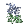 6lb7C 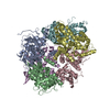 4nscS 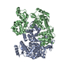 6iihS S: Starting model for refinement C: citing same article ( |
|---|---|
| Similar structure data |


-Links
 PDBj
PDBj


- Assembly
| Deposited unit | 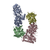
| ||||||||
|---|---|---|---|---|---|---|---|---|---|
| 1 | 
| ||||||||
| 2 | 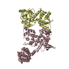
| ||||||||
| Unit cell |
|
-Components
| #1: Protein | Mass: 60321.707 Da / Num. of mol.: 2 / Mutation: C54T,C97A Source method: isolated from a genetically manipulated source Details: The fusion protein of T4L Endolysin, Linker, and Calcium uptake protein 1 Source: (gene. exp.) Escherichia virus T4, (gene. exp.) Homo sapiens (human)Gene: e, T4Tp126, MICU1, CALC, CBARA1 / Plasmid: pET-28b(+) / Production host:  #2: Protein | Mass: 38719.238 Da / Num. of mol.: 2 Source method: isolated from a genetically manipulated source Source: (gene. exp.) Homo sapiens (human) / Gene: MICU2, EFHA1 / Plasmid: pET-28b(+) / Production host: |
|---|
-Experimental details
-Experiment
| Experiment | Method: X-RAY DIFFRACTION / Number of used crystals: 1 |
|---|
- Sample preparation
| Crystal | Density Matthews: 2.38 Å3/Da / Density % sol: 48.24 % |
|---|---|
| Crystal grow | Temperature: 293.15 K / Method: vapor diffusion, hanging drop / pH: 5 Details: 2% tacsimate pH 5.0, 0.1 M sodium citrate pH 5.0, 12% (w/v) PEG 3350 |
-Data collection
| Diffraction | Mean temperature: 100 K / Serial crystal experiment: N |
|---|---|
| Diffraction source | Source: SYNCHROTRON / Site: SSRF / Beamline: BL19U1 / Wavelength: 0.97892 Å |
| Detector | Type: DECTRIS PILATUS3 S 6M / Detector: PIXEL / Date: Mar 7, 2019 |
| Radiation | Protocol: SINGLE WAVELENGTH / Monochromatic (M) / Laue (L): M / Scattering type: x-ray |
| Radiation wavelength | Wavelength: 0.97892 Å / Relative weight: 1 |
| Reflection | Resolution: 3.283→49.06 Å / Num. obs: 29560 / % possible obs: 99 % / Redundancy: 13 % / CC1/2: 0.958 / Rmerge(I) obs: 0.1947 / Rrim(I) all: 0.2028 / Net I/σ(I): 15.12 |
| Reflection shell | Resolution: 3.283→3.401 Å / Redundancy: 12.9 % / Rmerge(I) obs: 0.505 / Num. unique obs: 2680 / CC1/2: 0.937 / Rrim(I) all: 0.5255 / % possible all: 100 |
- Processing
| Software |
| ||||||||||||||||||||||||||||||||||||||||||||||||||||||||||||||||||||||||||||||||||||||||||
|---|---|---|---|---|---|---|---|---|---|---|---|---|---|---|---|---|---|---|---|---|---|---|---|---|---|---|---|---|---|---|---|---|---|---|---|---|---|---|---|---|---|---|---|---|---|---|---|---|---|---|---|---|---|---|---|---|---|---|---|---|---|---|---|---|---|---|---|---|---|---|---|---|---|---|---|---|---|---|---|---|---|---|---|---|---|---|---|---|---|---|---|
| Refinement | Method to determine structure: MOLECULAR REPLACEMENT Starting model: 4NSC, 6IIH Resolution: 3.283→49.056 Å / SU ML: 0.35 / Cross valid method: THROUGHOUT / σ(F): 1.34 / Phase error: 24.33 / Stereochemistry target values: ML
| ||||||||||||||||||||||||||||||||||||||||||||||||||||||||||||||||||||||||||||||||||||||||||
| Solvent computation | Shrinkage radii: 0.9 Å / VDW probe radii: 1.11 Å / Solvent model: FLAT BULK SOLVENT MODEL | ||||||||||||||||||||||||||||||||||||||||||||||||||||||||||||||||||||||||||||||||||||||||||
| Displacement parameters | Biso max: 128.61 Å2 / Biso mean: 39.8777 Å2 / Biso min: 4.94 Å2 | ||||||||||||||||||||||||||||||||||||||||||||||||||||||||||||||||||||||||||||||||||||||||||
| Refinement step | Cycle: final / Resolution: 3.283→49.056 Å
| ||||||||||||||||||||||||||||||||||||||||||||||||||||||||||||||||||||||||||||||||||||||||||
| Refine LS restraints |
| ||||||||||||||||||||||||||||||||||||||||||||||||||||||||||||||||||||||||||||||||||||||||||
| LS refinement shell | Refine-ID: X-RAY DIFFRACTION / Rfactor Rfree error: 0
|
