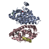
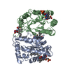
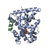
Entry Database : PDB / ID : 6lb5Title Crystal structure of dimeric RXR-LBD complexed with full agonist NEt-3IB and TIF2 co-activator Nuclear receptor coactivator 2 Retinoic acid receptor RXR-alpha Keywords / / / / / Function / homology Function Domain/homology Component
/ / / / / / / / / / / / / / / / / / / / / / / / / / / / / / / / / / / / / / / / / / / / / / / / / / / / / / / / / / / / / / / / / / / / / / / / / / / / / / / / / / / / / / / / / / / / / / / / / / / / / / / / / / / / / / / / / / / / / / / / / / / / / / / / / / / / / / / / / / / / / / / / / / / / / / / / / / / / / / / Biological species Homo sapiens (human)Method / / / Resolution : 2.4 Å Authors Imai, D. / Numoto, N. / Nakano, S. / Kakuta, H. / Ito, N. Journal : Biochem.Biophys.Res.Commun. / Year : 2024Title : Structural basis for the full and partial agonist activities of retinoid X receptor alpha ligands with an iso-butoxy and an isopropyl groupAuthors : Imai, D. / Numoto, N. / Tokiwa, H. / Kakuta, H. / Ito, N. History Deposition Nov 13, 2019 Deposition site / Processing site Revision 1.0 Nov 18, 2020 Provider / Type Revision 1.1 Nov 25, 2020 Group Category / pdbx_struct_assembly_gen / pdbx_struct_assembly_propRevision 1.2 Nov 22, 2023 Group / Database references / Refinement descriptionCategory chem_comp_atom / chem_comp_bond ... chem_comp_atom / chem_comp_bond / database_2 / pdbx_initial_refinement_model Item / _database_2.pdbx_database_accessionRevision 1.3 Oct 2, 2024 Group / Category / citation_authorItem _citation.country / _citation.journal_abbrev ... _citation.country / _citation.journal_abbrev / _citation.journal_id_ASTM / _citation.journal_id_CSD / _citation.journal_id_ISSN / _citation.journal_volume / _citation.page_first / _citation.pdbx_database_id_DOI / _citation.title / _citation.year / _citation_author.name
Show all Show less
 Movie
Movie Controller

 Yorodumi
 Open data
 Basic information
 Components
 Keywords
 Function and homology information
 Homo sapiens (human)
 X-RAY DIFFRACTION /
  Authors
 Citation
 Structure visualization
 Downloads & links
 Other downloads


 PDBj
PDBj


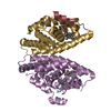
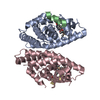
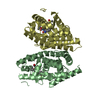
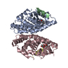
 Assembly
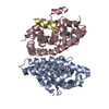

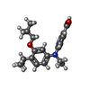
 Mass: 356.459 Da / Num. of mol.: 2 / Source method: obtained synthetically / Formula: C21H28N2O3 / Feature type: SUBJECT OF INVESTIGATION
 Mass: 18.015 Da / Num. of mol.: 165 / Source method: isolated from a natural source / Formula: H2O
 Sample preparation
 / Beamline: BL38B1 / Wavelength: 1 Å
 Processing