 Movie
Movie Controller

[English] 日本語
 Yorodumi
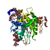
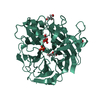
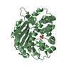
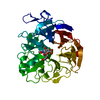
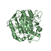
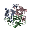
- PDB-6l8p: Crystal structure of RidA from Antarctic bacterium Psychrobacter ... -
+ Open data

- Basic information
| Entry | Database: PDB / ID: 6l8p | ||||||
|---|---|---|---|---|---|---|---|
| Title | Crystal structure of RidA from Antarctic bacterium Psychrobacter sp. PAMC 21119 | ||||||
 Components Components | RidA family protein | ||||||
 Keywords Keywords | HYDROLASE / RidA / Deamination / Psychrophile / Antarctic bacterium / Cold-adaptability | ||||||
| Function / homology |  Function and homology information Function and homology informationRidA, conserved site / Uncharacterized protein family UPF0076 signature. / RidA family / RutC-like / YjgF/YER057c/UK114 family / Endoribonuclease L-PSP / RutC-like superfamily / 60s Ribosomal Protein L30; Chain: A; / 2-Layer Sandwich / Alpha Beta Similarity search - Domain/homology | ||||||
| Biological species |  Psychrobacter sp. MES7-P7E (bacteria) Psychrobacter sp. MES7-P7E (bacteria) | ||||||
| Method |  X-RAY DIFFRACTION / SYNCHROTRON / MOLECULAR REPLACEMENT / Resolution: 1.601 Å X-RAY DIFFRACTION / SYNCHROTRON / MOLECULAR REPLACEMENT / Resolution: 1.601 Å | ||||||
 Authors Authors | Kwon, S. / Lee, C.W. / Koh, H.Y. / Lee, J.H. / Park, H.H. | ||||||
 Citation Citation | Journal: Biochem.Biophys.Res.Commun. / Year: 2020 Title: Crystal structure of the reactive intermediate/imine deaminase A homolog from the Antarctic bacterium Psychrobacter sp. PAMC 21119. Authors: Kwon, S. / Lee, C.W. / Koh, H.Y. / Park, H. / Lee, J.H. / Park, H.H. | ||||||
| History |
|
- Structure visualization
| Structure viewer | Molecule: MolmilJmol/JSmol |
|---|
- Downloads & links
-Download
| PDBx/mmCIF format | 6l8p.cif.gz | 94.6 KB | Display | PDBx/mmCIF format |
|---|---|---|---|---|
| PDB format | pdb6l8p.ent.gz | 69.2 KB | Display | PDB format |
| PDBx/mmJSON format | 6l8p.json.gz | Tree view | PDBx/mmJSON format | |
| Others |  Other downloads Other downloads |
-Validation report
| Summary document | 6l8p_validation.pdf.gz | 1.2 MB | Display | wwPDB validaton report |
|---|---|---|---|---|
| Full document | 6l8p_full_validation.pdf.gz | 1.2 MB | Display | |
| Data in XML | 6l8p_validation.xml.gz | 20 KB | Display | |
| Data in CIF | 6l8p_validation.cif.gz | 30.2 KB | Display | |
| Arichive directory | https://data.pdbj.org/pub/pdb/validation_reports/l8/6l8pftp://data.pdbj.org/pub/pdb/validation_reports/l8/6l8p | HTTPS FTP |
-Related structure data
| Related structure data | 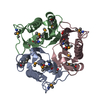 3k0tS S: Starting model for refinement |
|---|---|
| Similar structure data |


-Links
 PDBj
PDBj
- Assembly
| Deposited unit | 
| ||||||||
|---|---|---|---|---|---|---|---|---|---|
| 1 |
| ||||||||
| Unit cell |
|
-Components
| #1: Protein | Mass: 15665.569 Da / Num. of mol.: 3 / Mutation: D39E Source method: isolated from a genetically manipulated source Source: (gene. exp.) Psychrobacter sp. MES7-P7E (bacteria) / Gene: CXF62_02540 / Production host: #2: Chemical |   Mass: 102.046 Da / Num. of mol.: 3 / Source method: obtained synthetically / Formula: C3H2O4 / Feature type: SUBJECT OF INVESTIGATION Mass: 102.046 Da / Num. of mol.: 3 / Source method: obtained synthetically / Formula: C3H2O4 / Feature type: SUBJECT OF INVESTIGATION#3: Water | ChemComp-HOH / |  Mass: 18.015 Da / Num. of mol.: 460 / Source method: isolated from a natural source / Formula: H2O Mass: 18.015 Da / Num. of mol.: 460 / Source method: isolated from a natural source / Formula: H2OHas ligand of interest | Y | |
|---|
-Experimental details
-Experiment
| Experiment | Method: X-RAY DIFFRACTION / Number of used crystals: 1 |
|---|
- Sample preparation
| Crystal | Density Matthews: 2.76 Å3/Da / Density % sol: 55.47 % |
|---|---|
| Crystal grow | Temperature: 293 K / Method: vapor diffusion, hanging drop Details: 0.1 M Bis-Tris Propane/HCl (pH 7), 1.1 M malonic acid, 0.15 M ammonium citrate tribasic, 0.072 M succinic acid, 0.18 M DL-malic acid, 0.24 M sodium acetate, 0.3 M sodium formate, and 0.1 M ...Details: 0.1 M Bis-Tris Propane/HCl (pH 7), 1.1 M malonic acid, 0.15 M ammonium citrate tribasic, 0.072 M succinic acid, 0.18 M DL-malic acid, 0.24 M sodium acetate, 0.3 M sodium formate, and 0.1 M ammonium tartrate dibasic |
-Data collection
| Diffraction | Mean temperature: 100 K / Serial crystal experiment: N | |||||||||||||||||||||||||||||||||||||||||||||||||||||||||||||||||||||||||||||||||||||||||||||||||||||||||||||||||||||||||||||||||||||||||||||||||||||||||||||||||||||||||||||||||||||||||||||
|---|---|---|---|---|---|---|---|---|---|---|---|---|---|---|---|---|---|---|---|---|---|---|---|---|---|---|---|---|---|---|---|---|---|---|---|---|---|---|---|---|---|---|---|---|---|---|---|---|---|---|---|---|---|---|---|---|---|---|---|---|---|---|---|---|---|---|---|---|---|---|---|---|---|---|---|---|---|---|---|---|---|---|---|---|---|---|---|---|---|---|---|---|---|---|---|---|---|---|---|---|---|---|---|---|---|---|---|---|---|---|---|---|---|---|---|---|---|---|---|---|---|---|---|---|---|---|---|---|---|---|---|---|---|---|---|---|---|---|---|---|---|---|---|---|---|---|---|---|---|---|---|---|---|---|---|---|---|---|---|---|---|---|---|---|---|---|---|---|---|---|---|---|---|---|---|---|---|---|---|---|---|---|---|---|---|---|---|---|---|---|
| Diffraction source | Source: SYNCHROTRON / Site: PAL/PLS  / Beamline: 5C (4A) / Wavelength: 1 Å / Beamline: 5C (4A) / Wavelength: 1 Å | |||||||||||||||||||||||||||||||||||||||||||||||||||||||||||||||||||||||||||||||||||||||||||||||||||||||||||||||||||||||||||||||||||||||||||||||||||||||||||||||||||||||||||||||||||||||||||||
| Detector | Type: DECTRIS EIGER X 9M / Detector: PIXEL / Date: Apr 24, 2019 | |||||||||||||||||||||||||||||||||||||||||||||||||||||||||||||||||||||||||||||||||||||||||||||||||||||||||||||||||||||||||||||||||||||||||||||||||||||||||||||||||||||||||||||||||||||||||||||
| Radiation | Protocol: SINGLE WAVELENGTH / Monochromatic (M) / Laue (L): M / Scattering type: x-ray | |||||||||||||||||||||||||||||||||||||||||||||||||||||||||||||||||||||||||||||||||||||||||||||||||||||||||||||||||||||||||||||||||||||||||||||||||||||||||||||||||||||||||||||||||||||||||||||
| Radiation wavelength | Wavelength: 1 Å / Relative weight: 1 | |||||||||||||||||||||||||||||||||||||||||||||||||||||||||||||||||||||||||||||||||||||||||||||||||||||||||||||||||||||||||||||||||||||||||||||||||||||||||||||||||||||||||||||||||||||||||||||
| Reflection | Resolution: 1.6→50 Å / Num. obs: 69280 / % possible obs: 100 % / Redundancy: 6.9 % / Rmerge(I) obs: 0.106 / Rpim(I) all: 0.044 / Rrim(I) all: 0.115 / Χ2: 2.494 / Net I/σ(I): 10.6 / Num. measured all: 479686 | |||||||||||||||||||||||||||||||||||||||||||||||||||||||||||||||||||||||||||||||||||||||||||||||||||||||||||||||||||||||||||||||||||||||||||||||||||||||||||||||||||||||||||||||||||||||||||||
| Reflection shell | Diffraction-ID: 1
|
- Processing
| Software |
| ||||||||||||||||||||||||||||||||||||||||||||||||||||||||||||||||||||||||||||||||||||||||||
|---|---|---|---|---|---|---|---|---|---|---|---|---|---|---|---|---|---|---|---|---|---|---|---|---|---|---|---|---|---|---|---|---|---|---|---|---|---|---|---|---|---|---|---|---|---|---|---|---|---|---|---|---|---|---|---|---|---|---|---|---|---|---|---|---|---|---|---|---|---|---|---|---|---|---|---|---|---|---|---|---|---|---|---|---|---|---|---|---|---|---|---|
| Refinement | Method to determine structure: MOLECULAR REPLACEMENT Starting model: 3K0T Resolution: 1.601→42.227 Å / SU ML: 0.12 / Cross valid method: THROUGHOUT / σ(F): 1.37 / Phase error: 14.6
| ||||||||||||||||||||||||||||||||||||||||||||||||||||||||||||||||||||||||||||||||||||||||||
| Solvent computation | Shrinkage radii: 0.9 Å / VDW probe radii: 1.11 Å | ||||||||||||||||||||||||||||||||||||||||||||||||||||||||||||||||||||||||||||||||||||||||||
| Displacement parameters | Biso max: 79.17 Å2 / Biso mean: 17.355 Å2 / Biso min: 5.2 Å2 | ||||||||||||||||||||||||||||||||||||||||||||||||||||||||||||||||||||||||||||||||||||||||||
| Refinement step | Cycle: final / Resolution: 1.601→42.227 Å
| ||||||||||||||||||||||||||||||||||||||||||||||||||||||||||||||||||||||||||||||||||||||||||
| LS refinement shell | Refine-ID: X-RAY DIFFRACTION / Rfactor Rfree error: 0
|
