 Movie
Movie Controller


+ Open data

- Basic information
| Entry | Database: PDB / ID: 6l7i | ||||||
|---|---|---|---|---|---|---|---|
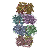
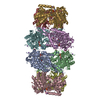
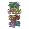
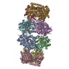
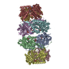
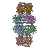
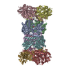
| Title | Signal substraction of TcdB1-TccC2 and part of TcdA1 | ||||||
 Components Components |
| ||||||
 Keywords Keywords | TOXIN/PROTEIN TRANSPORT / protein transportation / TcdA1 / TcdB1 / TccC2 / TOXIN-PROTEIN TRANSPORT complex | ||||||
| Function / homology |  Function and homology information Function and homology information | ||||||
| Biological species |  Photorhabdus luminescens (bacteria) Photorhabdus luminescens (bacteria) | ||||||
| Method | ELECTRON MICROSCOPY / single particle reconstruction / cryo EM / Resolution: 2.9 Å | ||||||
 Authors Authors | Yan, Z.F. / Yang, G.W. | ||||||
 Citation Citation | Journal: To Be Published Title: Signal substraction of TcdB1-TccC2 and part of TcdA1 Authors: Yan, Z.F. / Zhang, N. / Yang, G.W. | ||||||
| History |
|
- Structure visualization
| Movie |
Movie viewer |
|---|---|
| Structure viewer | Molecule: MolmilJmol/JSmol |
- Downloads & links
-Download
| PDBx/mmCIF format | 6l7i.cif.gz | 521.9 KB | Display | PDBx/mmCIF format |
|---|---|---|---|---|
| PDB format | pdb6l7i.ent.gz | 423.9 KB | Display | PDB format |
| PDBx/mmJSON format | 6l7i.json.gz | Tree view | PDBx/mmJSON format | |
| Others |  Other downloads Other downloads |
-Validation report
| Arichive directory | https://data.pdbj.org/pub/pdb/validation_reports/l7/6l7iftp://data.pdbj.org/pub/pdb/validation_reports/l7/6l7i | HTTPS FTP |
|---|
-Related structure data
| Related structure data | 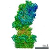 0844MC M: map data used to model this data C: citing same article ( |
|---|---|
| Similar structure data |

-Links
 PDBj
PDBj
- Assembly
| Deposited unit | 
|
|---|---|
| 1 |
|
-Components
| #1: Protein | Mass: 20255.887 Da / Num. of mol.: 5 Source method: isolated from a genetically manipulated source Source: (gene. exp.) Photorhabdus luminescens (bacteria) / Gene: tcdA, tcdA1 / Production host: #2: Protein | | Mass: 165296.453 Da / Num. of mol.: 1 Source method: isolated from a genetically manipulated source Source: (gene. exp.) Photorhabdus luminescens (bacteria) / Gene: tcdB1 / Production host: #3: Protein | | Mass: 74759.125 Da / Num. of mol.: 1 / Fragment: N terminus Source method: isolated from a genetically manipulated source Source: (gene. exp.) Photorhabdus luminescens (bacteria) / Gene: TccC2 / Production host: #4: Protein/peptide | | Mass: 1905.087 Da / Num. of mol.: 1 Source method: isolated from a genetically manipulated source Source: (gene. exp.) Photorhabdus luminescens (bacteria) / Gene: TccC2 / Production host: Has protein modification | Y | |
|---|
-Experimental details
-Experiment
| Experiment | Method: ELECTRON MICROSCOPY |
|---|---|
| EM experiment | Aggregation state: PARTICLE / 3D reconstruction method: single particle reconstruction |
- Sample preparation
| Component | Name: Toxin complex TcdA1-TcdB1-TccC2 / Type: COMPLEX / Entity ID: all / Source: RECOMBINANT |
|---|---|
| Source (natural) | Organism: Photorhabdus luminescens (bacteria) |
| Source (recombinant) | Organism: |
| Buffer solution | pH: 8 |
| Specimen | Embedding applied: NO / Shadowing applied: NO / Staining applied: NO / Vitrification applied: YES |
| Vitrification | Cryogen name: ETHANE |
- Electron microscopy imaging
| Microscopy | Model: FEI/PHILIPS CM300FEG/HE |
|---|---|
| Electron gun | Electron source:  FIELD EMISSION GUN / Accelerating voltage: 300 kV / Illumination mode: SPOT SCAN FIELD EMISSION GUN / Accelerating voltage: 300 kV / Illumination mode: SPOT SCAN |
| Electron lens | Mode: BRIGHT FIELD |
| Image recording | Electron dose: 50 e/Å2 / Film or detector model: GATAN K2 SUMMIT (4k x 4k) |
- Processing
| CTF correction | Type: NONE |
|---|---|
| 3D reconstruction | Resolution: 2.9 Å / Resolution method: FSC 0.143 CUT-OFF / Num. of particles: 124828 / Symmetry type: POINT |
