 Movie
Movie Controller

[English] 日本語
 Yorodumi
- PDB-6l5v: Carbonmonoxy human hemoglobin C in the R quaternary structure at ... -
+ Open data

- Basic information
| Entry | Database: PDB / ID: 6l5v | ||||||
|---|---|---|---|---|---|---|---|
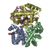
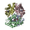
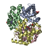
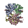
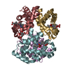
| Title | Carbonmonoxy human hemoglobin C in the R quaternary structure at 95 K: Light (2 min) | ||||||
 Components Components |
| ||||||
 Keywords Keywords | OXYGEN TRANSPORT / Hemoglobin / Photolysis | ||||||
| Function / homology |  Function and homology information Function and homology informationnitric oxide transport / hemoglobin binding / hemoglobin alpha binding / haptoglobin binding / haptoglobin-hemoglobin complex / organic acid binding / renal absorption / hemoglobin complex / oxygen transport / Scavenging of heme from plasma ...nitric oxide transport / hemoglobin binding / hemoglobin alpha binding / haptoglobin binding / haptoglobin-hemoglobin complex / organic acid binding / renal absorption / hemoglobin complex / oxygen transport / Scavenging of heme from plasma / endocytic vesicle lumen / blood vessel diameter maintenance / hydrogen peroxide catabolic process / oxygen carrier activity / Late endosomal microautophagy / Heme signaling / carbon dioxide transport / response to hydrogen peroxide / Erythrocytes take up oxygen and release carbon dioxide / Erythrocytes take up carbon dioxide and release oxygen / Cytoprotection by HMOX1 / oxygen binding / regulation of blood pressure / platelet aggregation / Chaperone Mediated Autophagy / peroxidase activity / positive regulation of nitric oxide biosynthetic process / tertiary granule lumen / Factors involved in megakaryocyte development and platelet production / ficolin-1-rich granule lumen / blood microparticle / iron ion binding / heme binding / Neutrophil degranulation / extracellular space / extracellular exosome / extracellular region / membrane / metal ion binding / cytosol Similarity search - Function | ||||||
| Biological species |  Homo sapiens (human) Homo sapiens (human) | ||||||
| Method |  X-RAY DIFFRACTION / SYNCHROTRON / MOLECULAR REPLACEMENT / Resolution: 1.45 Å X-RAY DIFFRACTION / SYNCHROTRON / MOLECULAR REPLACEMENT / Resolution: 1.45 Å | ||||||
 Authors Authors | Shibayama, N. / Park, S.Y. / Ohki, M. / Sato-Tomita, A. | ||||||
| Funding support |  Japan, 1items Japan, 1items
| ||||||
 Citation Citation | Journal: Proc.Natl.Acad.Sci.USA / Year: 2020 Title: Direct observation of ligand migration within human hemoglobin at work. Authors: Shibayama, N. / Sato-Tomita, A. / Ohki, M. / Ichiyanagi, K. / Park, S.Y. | ||||||
| History |
|
- Structure visualization
| Structure viewer | Molecule: MolmilJmol/JSmol |
|---|
- Downloads & links
-Download
| PDBx/mmCIF format | 6l5v.cif.gz | 134.5 KB | Display | PDBx/mmCIF format |
|---|---|---|---|---|
| PDB format | pdb6l5v.ent.gz | 103.1 KB | Display | PDB format |
| PDBx/mmJSON format | 6l5v.json.gz | Tree view | PDBx/mmJSON format | |
| Others |  Other downloads Other downloads |
-Validation report
| Summary document | 6l5v_validation.pdf.gz | 1.1 MB | Display | wwPDB validaton report |
|---|---|---|---|---|
| Full document | 6l5v_full_validation.pdf.gz | 1.1 MB | Display | |
| Data in XML | 6l5v_validation.xml.gz | 14.4 KB | Display | |
| Data in CIF | 6l5v_validation.cif.gz | 19.3 KB | Display | |
| Arichive directory | https://data.pdbj.org/pub/pdb/validation_reports/l5/6l5vftp://data.pdbj.org/pub/pdb/validation_reports/l5/6l5v | HTTPS FTP |
-Related structure data
| Related structure data |  6ka9C  6kaeC  6kahC  6kaiC  6kaoC  6kapC  6kaqC  6karC  6kasC  6katC  6kauC  6kavC  6l5wC  6l5xC  6l5yC  6lcwC  6lcxC  3s66S C: citing same article ( S: Starting model for refinement |
|---|---|
| Similar structure data |

-Links
 PDBj
PDBj


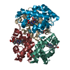
- Assembly
| Deposited unit | 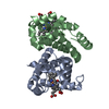
| ||||||||
|---|---|---|---|---|---|---|---|---|---|
| 1 | 
| ||||||||
| Unit cell |
|
-Components
| #1: Protein | Mass: 15150.353 Da / Num. of mol.: 1 / Source method: isolated from a natural source / Source: (natural) Homo sapiens (human) / References: UniProt: P69905 | ||||||
|---|---|---|---|---|---|---|---|
| #2: Protein | Mass: 15890.265 Da / Num. of mol.: 1 / Source method: isolated from a natural source / Source: (natural) Homo sapiens (human) / References: UniProt: P68871 | ||||||
| #3: Chemical |   Mass: 616.487 Da / Num. of mol.: 2 / Source method: obtained synthetically / Formula: C34H32FeN4O4 Mass: 616.487 Da / Num. of mol.: 2 / Source method: obtained synthetically / Formula: C34H32FeN4O4#4: Chemical |   Mass: 28.010 Da / Num. of mol.: 2 / Source method: obtained synthetically / Formula: CO Mass: 28.010 Da / Num. of mol.: 2 / Source method: obtained synthetically / Formula: CO#5: Water | ChemComp-HOH / |  Mass: 18.015 Da / Num. of mol.: 122 / Source method: isolated from a natural source / Formula: H2O Mass: 18.015 Da / Num. of mol.: 122 / Source method: isolated from a natural source / Formula: H2OHas ligand of interest | N | |
-Experimental details
-Experiment
| Experiment | Method: X-RAY DIFFRACTION / Number of used crystals: 1 |
|---|
- Sample preparation
| Crystal | Density Matthews: 2.22 Å3/Da / Density % sol: 44.61 % |
|---|---|
| Crystal grow | Temperature: 293 K / Method: batch mode / pH: 7.2 Details: 0.528 M sodium dihydrogen phosphate, 1.122 M dipotassium hydrogen phosphate, 10%v/v glycerol |
-Data collection
| Diffraction | Mean temperature: 95 K / Serial crystal experiment: N | ||||||||||||||||||||||||||||||
|---|---|---|---|---|---|---|---|---|---|---|---|---|---|---|---|---|---|---|---|---|---|---|---|---|---|---|---|---|---|---|---|
| Diffraction source | Source: SYNCHROTRON / Site: Photon Factory / Beamline: AR-NW12A / Wavelength: 1 Å | ||||||||||||||||||||||||||||||
| Detector | Type: DECTRIS PILATUS3 S 2M / Detector: PIXEL / Date: Nov 23, 2018 | ||||||||||||||||||||||||||||||
| Radiation | Protocol: SINGLE WAVELENGTH / Monochromatic (M) / Laue (L): M / Scattering type: x-ray | ||||||||||||||||||||||||||||||
| Radiation wavelength | Wavelength: 1 Å / Relative weight: 1 | ||||||||||||||||||||||||||||||
| Reflection | Resolution: 1.45→18.91 Å / Num. obs: 50700 / % possible obs: 99.8 % / Redundancy: 11.1 % / Biso Wilson estimate: 17.85 Å2 / CC1/2: 0.999 / Rmerge(I) obs: 0.066 / Rpim(I) all: 0.021 / Rrim(I) all: 0.069 / Net I/σ(I): 19.1 | ||||||||||||||||||||||||||||||
| Reflection shell | Diffraction-ID: 1
|
- Processing
| Software |
| ||||||||||||||||||||||||||||||||||||||||||||||||||||||||||||||||||||||||||||||||||||||||||||||||||||||||||||||||||
|---|---|---|---|---|---|---|---|---|---|---|---|---|---|---|---|---|---|---|---|---|---|---|---|---|---|---|---|---|---|---|---|---|---|---|---|---|---|---|---|---|---|---|---|---|---|---|---|---|---|---|---|---|---|---|---|---|---|---|---|---|---|---|---|---|---|---|---|---|---|---|---|---|---|---|---|---|---|---|---|---|---|---|---|---|---|---|---|---|---|---|---|---|---|---|---|---|---|---|---|---|---|---|---|---|---|---|---|---|---|---|---|---|---|---|---|
| Refinement | Method to determine structure: MOLECULAR REPLACEMENT Starting model: 3S66 Resolution: 1.45→18.91 Å / SU ML: 0.12 / Cross valid method: THROUGHOUT / σ(F): 1.34 / Phase error: 19.79
| ||||||||||||||||||||||||||||||||||||||||||||||||||||||||||||||||||||||||||||||||||||||||||||||||||||||||||||||||||
| Solvent computation | Shrinkage radii: 0.9 Å / VDW probe radii: 1.11 Å | ||||||||||||||||||||||||||||||||||||||||||||||||||||||||||||||||||||||||||||||||||||||||||||||||||||||||||||||||||
| Displacement parameters | Biso max: 112.54 Å2 / Biso mean: 27.9434 Å2 / Biso min: 10.53 Å2 | ||||||||||||||||||||||||||||||||||||||||||||||||||||||||||||||||||||||||||||||||||||||||||||||||||||||||||||||||||
| Refinement step | Cycle: final / Resolution: 1.45→18.91 Å
| ||||||||||||||||||||||||||||||||||||||||||||||||||||||||||||||||||||||||||||||||||||||||||||||||||||||||||||||||||
| Refine LS restraints |
| ||||||||||||||||||||||||||||||||||||||||||||||||||||||||||||||||||||||||||||||||||||||||||||||||||||||||||||||||||
| LS refinement shell | Refine-ID: X-RAY DIFFRACTION / Rfactor Rfree error: 0
| ||||||||||||||||||||||||||||||||||||||||||||||||||||||||||||||||||||||||||||||||||||||||||||||||||||||||||||||||||
| Refinement TLS params. | Method: refined / Origin x: 23.6506 Å / Origin y: -19.7633 Å / Origin z: 11.302 Å
| ||||||||||||||||||||||||||||||||||||||||||||||||||||||||||||||||||||||||||||||||||||||||||||||||||||||||||||||||||
| Refinement TLS group | Selection details: all |
