- PDB-6l4p: Crystal structure of the complex between the axonemal outer-arm d... -
+
Open data
ID or keywords:
Loading...
-
Basic information
Entry
Database: PDB / ID: 6l4p
Title
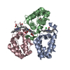
Crystal structure of the complex between the axonemal outer-arm dynein light chain-1 and microtubule binding domain of gamma heavy chain
Components
Dynein light chain 1, axonemal
Flagellar outer dynein arm heavy chain gamma
Keywords
CONTRACTILE PROTEIN/MOTOR PROTEIN / Motor protein / Complex / CONTRACTILE PROTEIN-MOTOR PROTEIN complex
Function / homology
Function and homology information
outer dynein arm / outer dynein arm assembly / cilium movement involved in cell motility / dynein complex / motile cilium / minus-end-directed microtubule motor activity / dynein light intermediate chain binding / microtubule-based movement / dynein intermediate chain binding / microtubule ...outer dynein arm / outer dynein arm assembly / cilium movement involved in cell motility / dynein complex / motile cilium / minus-end-directed microtubule motor activity / dynein light intermediate chain binding / microtubule-based movement / dynein intermediate chain binding / microtubule / ATP binding / cytoplasm Similarity search - Function
: / Dynein axonemal heavy chain 2/5/8 coiled-coil / Dynein heavy chain 3, AAA+ lid domain / AAA+ lid domain / Leucine rich repeat 4 / Leucine Rich repeats (2 copies) / Dynein heavy chain, AAA 5 extension domain / Dynein heavy chain AAA lid domain / Dynein heavy chain, C-terminal domain / Dynein heavy chain, C-terminal domain, barrel region ...: / Dynein axonemal heavy chain 2/5/8 coiled-coil / Dynein heavy chain 3, AAA+ lid domain / AAA+ lid domain / Leucine rich repeat 4 / Leucine Rich repeats (2 copies) / Dynein heavy chain, AAA 5 extension domain / Dynein heavy chain AAA lid domain / Dynein heavy chain, C-terminal domain / Dynein heavy chain, C-terminal domain, barrel region / Dynein heavy chain C-terminal domain / Dynein heavy chain, tail / Dynein heavy chain, N-terminal region 1 / P-loop containing dynein motor region / Dynein heavy chain region D6 P-loop domain / Dynein heavy chain, linker / Dynein heavy chain, AAA module D4 / Dynein heavy chain, coiled coil stalk / Dynein heavy chain / Dynein heavy chain, hydrolytic ATP-binding dynein motor region / Dynein heavy chain, ATP-binding dynein motor region / Dynein heavy chain AAA lid domain / Dynein heavy chain AAA lid domain superfamily / Dynein heavy chain, domain 2, N-terminal / Dynein heavy chain, linker, subdomain 3 / Dynein heavy chain, AAA1 domain, small subdomain / Dynein heavy chain region D6 P-loop domain / Dynein heavy chain, N-terminal region 2 / Hydrolytic ATP binding site of dynein motor region / Microtubule-binding stalk of dynein motor / P-loop containing dynein motor region D4 / ATP-binding dynein motor region / Dynein heavy chain AAA lid domain / Leucine-rich repeat, SDS22-like subfamily / Leucine-rich repeat profile. / Leucine-rich repeat / Leucine-rich repeat domain superfamily / ATPases associated with a variety of cellular activities / AAA+ ATPase domain / P-loop containing nucleoside triphosphate hydrolase Similarity search - Domain/homology
PHOSPHATE ION / Flagellar outer dynein arm heavy chain gamma / Dynein gamma chain, flagellar outer arm / Dynein axonemal light chain 1 Similarity search - Component
In the structure databanks used in Yorodumi, some data are registered as the other names, "COVID-19 virus" and "2019-nCoV". Here are the details of the virus and the list of structure data.
Jan 31, 2019. EMDB accession codes are about to change! (news from PDBe EMDB page)
EMDB accession codes are about to change! (news from PDBe EMDB page)
The allocation of 4 digits for EMDB accession codes will soon come to an end. Whilst these codes will remain in use, new EMDB accession codes will include an additional digit and will expand incrementally as the available range of codes is exhausted. The current 4-digit format prefixed with “EMD-” (i.e. EMD-XXXX) will advance to a 5-digit format (i.e. EMD-XXXXX), and so on. It is currently estimated that the 4-digit codes will be depleted around Spring 2019, at which point the 5-digit format will come into force.
The EM Navigator/Yorodumi systems omit the EMD- prefix.
Related info.:Q: What is EMD? / ID/Accession-code notation in Yorodumi/EM Navigator
Yorodumi is a browser for structure data from EMDB, PDB, SASBDB, etc.
This page is also the successor to EM Navigator detail page, and also detail information page/front-end page for Omokage search.
The word "yorodu" (or yorozu) is an old Japanese word meaning "ten thousand". "mi" (miru) is to see.
Related info.:EMDB / PDB / SASBDB / Comparison of 3 databanks / Yorodumi Search / Aug 31, 2016. New EM Navigator & Yorodumi / Yorodumi Papers / Jmol/JSmol / Function and homology information / Changes in new EM Navigator and Yorodumi
 Movie
Movie Controller

 Yorodumi
 Open data
 Basic information
 Components
 Keywords
 Function and homology information
 Chlamydomonas reinhardtii (plant)
 X-RAY DIFFRACTION /
  Authors
 Japan, 2items
  Citation
 Structure visualization
 Downloads & links
 Other downloads
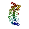


 PDBj
PDBj


 Assembly


 Mass: 94.971 Da / Num. of mol.: 1 / Source method: obtained synthetically / Formula: PO4
 Mass: 122.143 Da / Num. of mol.: 1 / Source method: obtained synthetically / Formula: C4H12NO3 / Comment: pH buffer*YM
 Mass: 22.990 Da / Num. of mol.: 1 / Source method: obtained synthetically / Formula: Na
 Sample preparation
 Processing