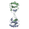
 Movie
Movie Controller


+ Open data

- Basic information
| Entry | Database: PDB / ID: 6l4n | ||||||
|---|---|---|---|---|---|---|---|
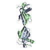
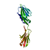
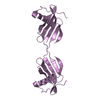
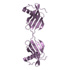
| Title | Domain swapped dimer of Monellin loop1 mutant with QVPAG motif | ||||||
 Components Components | Single chain Monellin | ||||||
 Keywords Keywords | PLANT PROTEIN / Monomer / L1 mutant | ||||||
| Biological species |  Dioscoreophyllum cumminsii (serendipity berry) Dioscoreophyllum cumminsii (serendipity berry) | ||||||
| Method |  X-RAY DIFFRACTION / MOLECULAR REPLACEMENT / Resolution: 2.431 Å X-RAY DIFFRACTION / MOLECULAR REPLACEMENT / Resolution: 2.431 Å | ||||||
 Authors Authors | Manjula, R. / Ramaswamy, S. / Gosavi, S. | ||||||
 Citation Citation | Journal: To Be Published Title: Domain swapped dimer of Monellin lopp1 mutant with QVPAG motif Authors: Manjula, R. / Ramaswamy, S. / Gosavi, S. | ||||||
| History |
|
- Structure visualization
| Structure viewer | Molecule:  MolmilJmol/JSmol MolmilJmol/JSmol |
|---|
- Downloads & links
-Download
| PDBx/mmCIF format | 6l4n.cif.gz | 52.8 KB | Display | PDBx/mmCIF format |
|---|---|---|---|---|
| PDB format | pdb6l4n.ent.gz | 35.9 KB | Display | PDB format |
| PDBx/mmJSON format | 6l4n.json.gz | Tree view | PDBx/mmJSON format | |
| Others |  Other downloads Other downloads |
-Validation report
| Arichive directory | https://data.pdbj.org/pub/pdb/validation_reports/l4/6l4nftp://data.pdbj.org/pub/pdb/validation_reports/l4/6l4n | HTTPS FTP |
|---|
-Related structure data
| Related structure data |  2o9aS S: Starting model for refinement |
|---|---|
| Similar structure data |


-Links
 PDBj
PDBj- Assembly
| Deposited unit | 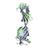
| |||||||||||||||||||||||||||||||||||||||||||||
|---|---|---|---|---|---|---|---|---|---|---|---|---|---|---|---|---|---|---|---|---|---|---|---|---|---|---|---|---|---|---|---|---|---|---|---|---|---|---|---|---|---|---|---|---|---|---|
| 1 |
| |||||||||||||||||||||||||||||||||||||||||||||
| Unit cell |
| |||||||||||||||||||||||||||||||||||||||||||||
| Noncrystallographic symmetry (NCS) | NCS domain:
NCS domain segments:
|
-Components
| #1: Protein | Mass: 10546.125 Da / Num. of mol.: 2 Source method: isolated from a genetically manipulated source Source: (gene. exp.) Dioscoreophyllum cumminsii (serendipity berry)Production host:  #2: Chemical | ChemComp-MG /   Mass: 24.305 Da / Num. of mol.: 4 / Source method: isolated from a natural source / Formula: Mg / Feature type: SUBJECT OF INVESTIGATION Mass: 24.305 Da / Num. of mol.: 4 / Source method: isolated from a natural source / Formula: Mg / Feature type: SUBJECT OF INVESTIGATION#3: Water | ChemComp-HOH / |  Mass: 18.015 Da / Num. of mol.: 55 / Source method: isolated from a natural source / Formula: H2O Mass: 18.015 Da / Num. of mol.: 55 / Source method: isolated from a natural source / Formula: H2OHas ligand of interest | Y | Sequence details | The complete sequence of single chain Monellin has been deposited to NCBI with accession code ...The complete sequence of single chain Monellin has been deposited to NCBI with accession code AFF58925. Residues 48-57 YENEGFREIK | |
|---|
-Experimental details
-Experiment
| Experiment | Method: X-RAY DIFFRACTION / Number of used crystals: 1 |
|---|
- Sample preparation
| Crystal | Density Matthews: 2.33 Å3/Da / Density % sol: 47.1 % |
|---|---|
| Crystal grow | Temperature: 293 K / Method: vapor diffusion, sitting drop / pH: 6.8 / Details: 20% PEG 3350, 200mM Sodium thiocyanate 7.0 |
-Data collection
| Diffraction | Mean temperature: 80 K / Serial crystal experiment: N | ||||||||||||||||||||||||||||||
|---|---|---|---|---|---|---|---|---|---|---|---|---|---|---|---|---|---|---|---|---|---|---|---|---|---|---|---|---|---|---|---|
| Diffraction source | Source: ROTATING ANODE / Type: RIGAKU FR-X / Wavelength: 1.5478 Å | ||||||||||||||||||||||||||||||
| Detector | Type: RIGAKU RAXIS IV++ / Detector: IMAGE PLATE / Date: May 21, 2019 | ||||||||||||||||||||||||||||||
| Radiation | Protocol: SINGLE WAVELENGTH / Monochromatic (M) / Laue (L): M / Scattering type: x-ray | ||||||||||||||||||||||||||||||
| Radiation wavelength | Wavelength: 1.5478 Å / Relative weight: 1 | ||||||||||||||||||||||||||||||
| Reflection | Resolution: 2.43→41.872 Å / Num. obs: 7283 / % possible obs: 93.4 % / Redundancy: 2.5 % / CC1/2: 0.995 / Rmerge(I) obs: 0.049 / Rpim(I) all: 0.037 / Rrim(I) all: 0.062 / Net I/σ(I): 11.1 | ||||||||||||||||||||||||||||||
| Reflection shell | Diffraction-ID: 1
|
- Processing
| Software |
| ||||||||||||||||||||||||
|---|---|---|---|---|---|---|---|---|---|---|---|---|---|---|---|---|---|---|---|---|---|---|---|---|---|
| Refinement | Method to determine structure: MOLECULAR REPLACEMENT Starting model: 2O9A Resolution: 2.431→41.87 Å / SU ML: 0.36 / Cross valid method: THROUGHOUT / σ(F): 1.39 / Phase error: 26.43
| ||||||||||||||||||||||||
| Solvent computation | Shrinkage radii: 0.9 Å / VDW probe radii: 1.11 Å | ||||||||||||||||||||||||
| Displacement parameters | Biso max: 79.24 Å2 / Biso mean: 35.5651 Å2 / Biso min: 16.48 Å2 | ||||||||||||||||||||||||
| Refinement step | Cycle: final / Resolution: 2.431→41.87 Å
| ||||||||||||||||||||||||
| Refine LS restraints NCS |
| ||||||||||||||||||||||||
| LS refinement shell | Refine-ID: X-RAY DIFFRACTION / Rfactor Rfree error: 0
|
