 Movie
Movie Controller

[English] 日本語
 Yorodumi
- PDB-6l3y: Crystal Structure of Lysyl-tRNA Synthetase from Plasmodium falcip... -
+ Open data

- Basic information
| Entry | Database: PDB / ID: 6l3y | ||||||
|---|---|---|---|---|---|---|---|
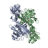
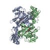
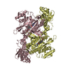
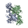
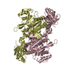
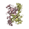
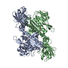
| Title | Crystal Structure of Lysyl-tRNA Synthetase from Plasmodium falciparum complexed with L-lysine and Clado-C | ||||||
 Components Components | Lysine--tRNA ligase | ||||||
 Keywords Keywords | PROTEIN BINDING/INHIBITOR / Inhibitor / Cladosporin analog / Stereochemistry / PROTEIN BINDING / PROTEIN BINDING-INHIBITOR complex | ||||||
| Function / homology |  Function and homology information Function and homology informationTranscriptional and post-translational regulation of MITF-M expression and activity / lysine-tRNA ligase / lysine-tRNA ligase activity / lysyl-tRNA aminoacylation / diadenosine tetraphosphate biosynthetic process / tRNA binding / ATP binding / cytoplasm Similarity search - Function | ||||||
| Biological species |  | ||||||
| Method |  X-RAY DIFFRACTION / SYNCHROTRON / MOLECULAR REPLACEMENT / Resolution: 3.1 Å X-RAY DIFFRACTION / SYNCHROTRON / MOLECULAR REPLACEMENT / Resolution: 3.1 Å | ||||||
 Authors Authors | Babbar, P. / Sharma, A. / Mishra, S. / Manickam, Y. / Harlos, K. | ||||||
| Funding support |  India, 1items India, 1items
| ||||||
 Citation Citation | Journal: Chembiochem / Year: 2021 Title: Crystal Structure of Lysyl-tRNA Synthetase from Plasmodium falciparum complexed with L-lysine and Cladosporin inhibitor, Cla-B Authors: Babbar, P. / Sato, M. / Manickam, Y. / Mishra, S. / Harlos, K. / Gupta, S. / Parvez, S. / Kikuchi, H. / Sharma, A. #1: Journal: J. Struct. Funct. Genomics / Year: 2014Title: Structural basis of malaria parasite lysyl-tRNA synthetase inhibition by cladosporin. Authors: Khan, S. / Sharma, A. / Belrhali, H. / Yogavel, M. / Sharma, A. | ||||||
| History |
|
- Structure visualization
| Structure viewer | Molecule: MolmilJmol/JSmol |
|---|
- Downloads & links
-Download
| PDBx/mmCIF format | 6l3y.cif.gz | 208.1 KB | Display | PDBx/mmCIF format |
|---|---|---|---|---|
| PDB format | pdb6l3y.ent.gz | 164.9 KB | Display | PDB format |
| PDBx/mmJSON format | 6l3y.json.gz | Tree view | PDBx/mmJSON format | |
| Others |  Other downloads Other downloads |
-Validation report
| Arichive directory | https://data.pdbj.org/pub/pdb/validation_reports/l3/6l3yftp://data.pdbj.org/pub/pdb/validation_reports/l3/6l3y | HTTPS FTP |
|---|
-Related structure data
| Related structure data |  6l4qC 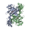 4pg3S S: Starting model for refinement C: citing same article ( |
|---|---|
| Similar structure data |


-Links
 PDBj
PDBj

- Assembly
| Deposited unit | 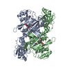
| ||||||||
|---|---|---|---|---|---|---|---|---|---|
| 1 |
| ||||||||
| Unit cell |
|
-Components
| #1: Protein | Mass: 58706.934 Da / Num. of mol.: 2 Source method: isolated from a genetically manipulated source Source: (gene. exp.)  #2: Chemical |   Type: L-peptide linking / Mass: 147.195 Da / Num. of mol.: 2 / Source method: obtained synthetically / Formula: C6H15N2O2 Type: L-peptide linking / Mass: 147.195 Da / Num. of mol.: 2 / Source method: obtained synthetically / Formula: C6H15N2O2#3: Chemical |   Mass: 305.369 Da / Num. of mol.: 2 / Source method: obtained synthetically / Formula: C17H23NO4 / Feature type: SUBJECT OF INVESTIGATION Mass: 305.369 Da / Num. of mol.: 2 / Source method: obtained synthetically / Formula: C17H23NO4 / Feature type: SUBJECT OF INVESTIGATION#4: Chemical | ChemComp-PO4 /   Mass: 94.971 Da / Num. of mol.: 5 / Source method: isolated from a natural source / Formula: PO4 Mass: 94.971 Da / Num. of mol.: 5 / Source method: isolated from a natural source / Formula: PO4#5: Water | ChemComp-HOH / |  Mass: 18.015 Da / Num. of mol.: 20 / Source method: isolated from a natural source / Formula: H2O Mass: 18.015 Da / Num. of mol.: 20 / Source method: isolated from a natural source / Formula: H2OHas ligand of interest | Y | Has protein modification | Y | |
|---|
-Experimental details
-Experiment
| Experiment | Method: X-RAY DIFFRACTION / Number of used crystals: 1 |
|---|
- Sample preparation
| Crystal | Density Matthews: 2.33 Å3/Da / Density % sol: 47.13 % Description: THE ENTRY CONTAINS FRIEDEL PAIRS IN I/F_PLUS/MINUS COLUMNS. |
|---|---|
| Crystal grow | Temperature: 293.15 K / Method: vapor diffusion, hanging drop Details: 0.09M NPS-0.3M Sodium nitrate, 0.3 Sodium phosphate dibasic,0.3M Ammonium sulfate. 1.0M Buffer-pH6.5- Imidazole; MES monohydrate (acid) Precipitant Mix-25% v/v MPD; 25% PEG 1000; 25% w/v PEG 3350 |
-Data collection
| Diffraction | Mean temperature: 293 K / Serial crystal experiment: N |
|---|---|
| Diffraction source | Source: SYNCHROTRON / Site: Diamond  / Beamline: I04 / Wavelength: 0.9795 Å / Beamline: I04 / Wavelength: 0.9795 Å |
| Detector | Type: DECTRIS EIGER X 16M / Detector: PIXEL / Date: May 6, 2019 |
| Radiation | Protocol: SINGLE WAVELENGTH / Monochromatic (M) / Laue (L): M / Scattering type: x-ray |
| Radiation wavelength | Wavelength: 0.9795 Å / Relative weight: 1 |
| Reflection | Resolution: 2.89→149.921 Å / Num. obs: 25891 / % possible obs: 100 % / Redundancy: 13.3 % / CC1/2: 0.989 / Net I/σ(I): 5.7 |
| Reflection shell | Resolution: 2.898→2.948 Å / Mean I/σ(I) obs: 0.8 / Num. unique obs: 1293 / CC1/2: 0.446 / % possible all: 100 |
- Processing
| Software |
| |||||||||||||||||||||||||||||||||||||||||||||
|---|---|---|---|---|---|---|---|---|---|---|---|---|---|---|---|---|---|---|---|---|---|---|---|---|---|---|---|---|---|---|---|---|---|---|---|---|---|---|---|---|---|---|---|---|---|---|
| Refinement | Method to determine structure: MOLECULAR REPLACEMENT Starting model: 4PG3 Resolution: 3.1→74.96 Å / SU ML: 0.37 / Cross valid method: THROUGHOUT / σ(F): 1.34 / Phase error: 30.32 / Stereochemistry target values: ML
| |||||||||||||||||||||||||||||||||||||||||||||
| Solvent computation | Shrinkage radii: 0.9 Å / VDW probe radii: 1.11 Å / Solvent model: FLAT BULK SOLVENT MODEL | |||||||||||||||||||||||||||||||||||||||||||||
| Displacement parameters | Biso max: 117.58 Å2 / Biso mean: 57.6733 Å2 / Biso min: 26.95 Å2 | |||||||||||||||||||||||||||||||||||||||||||||
| Refinement step | Cycle: final / Resolution: 3.1→74.96 Å
| |||||||||||||||||||||||||||||||||||||||||||||
| LS refinement shell | Refine-ID: X-RAY DIFFRACTION / Rfactor Rfree error: 0 / % reflection obs: 100 %
|
