 Movie
Movie Controller

[English] 日本語
 Yorodumi
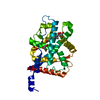
- PDB-6l2w: Crystal structure of a novel fold protein Gp72 from the freshwate... -
+ Open data

- Basic information
| Entry | Database: PDB / ID: 6l2w | ||||||||||||||||||
|---|---|---|---|---|---|---|---|---|---|---|---|---|---|---|---|---|---|---|---|
| Title | Crystal structure of a novel fold protein Gp72 from the freshwater cyanophage Mic1 | ||||||||||||||||||
 Components Components | freshwater cyanophage protein | ||||||||||||||||||
 Keywords Keywords | STRUCTURAL PROTEIN / hypothetical proteins | ||||||||||||||||||
| Function / homology | Uncharacterized protein Function and homology information Function and homology information | ||||||||||||||||||
| Biological species |  Microcystis phage Mic1 (virus) Microcystis phage Mic1 (virus) | ||||||||||||||||||
| Method |  X-RAY DIFFRACTION / SYNCHROTRON / SAD / Resolution: 2.29 Å X-RAY DIFFRACTION / SYNCHROTRON / SAD / Resolution: 2.29 Å | ||||||||||||||||||
 Authors Authors | Wang, Y. / Jin, H. / Yang, F. / Jiang, Y.L. / Zhao, Y.Y. / Chen, Z.P. / Li, W.F. / Chen, Y. / Zhou, C.Z. / Li, Q. | ||||||||||||||||||
| Funding support |  China, 5items China, 5items
| ||||||||||||||||||
 Citation Citation | Journal: Proteins / Year: 2020 Title: Crystal structure of a novel fold protein Gp72 from the freshwater cyanophage Mic1. Authors: Wang, Y. / Jin, H. / Yang, F. / Jiang, Y.L. / Zhao, Y.Y. / Chen, Z.P. / Li, W.F. / Chen, Y. / Zhou, C.Z. / Li, Q. | ||||||||||||||||||
| History |
|
- Structure visualization
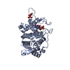
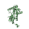
| Structure viewer | Molecule: MolmilJmol/JSmol |
|---|
- Downloads & links
-Download
| PDBx/mmCIF format | 6l2w.cif.gz | 61.9 KB | Display | PDBx/mmCIF format |
|---|---|---|---|---|
| PDB format | pdb6l2w.ent.gz | 45.1 KB | Display | PDB format |
| PDBx/mmJSON format | 6l2w.json.gz | Tree view | PDBx/mmJSON format | |
| Others |  Other downloads Other downloads |
-Validation report
| Arichive directory | https://data.pdbj.org/pub/pdb/validation_reports/l2/6l2wftp://data.pdbj.org/pub/pdb/validation_reports/l2/6l2w | HTTPS FTP |
|---|
-Related structure data
| Similar structure data |
|---|


-Links
 PDBj
PDBj- Assembly
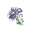
| Deposited unit | 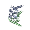
| ||||||||
|---|---|---|---|---|---|---|---|---|---|
| 1 |
| ||||||||
| Unit cell |
| ||||||||
| Components on special symmetry positions |
|
-Components
| #1: Protein | Mass: 14636.416 Da / Num. of mol.: 2 Source method: isolated from a genetically manipulated source Source: (gene. exp.) Microcystis phage Mic1 (virus) / Production host:  #2: Water | ChemComp-HOH / |  Mass: 18.015 Da / Num. of mol.: 57 / Source method: isolated from a natural source / Formula: H2O Mass: 18.015 Da / Num. of mol.: 57 / Source method: isolated from a natural source / Formula: H2O |
|---|
-Experimental details
-Experiment
| Experiment | Method: X-RAY DIFFRACTION / Number of used crystals: 1 |
|---|
- Sample preparation
| Crystal | Density Matthews: 2.24 Å3/Da / Density % sol: 45.06 % |
|---|---|
| Crystal grow | Temperature: 289 K / Method: vapor diffusion, sitting drop / pH: 7 Details: 13% polyethylene glycol 6000, 0.1 M ADA pH 7.0, 0.01 M spermine tetrahydrochloride and 0.1 M guanidine hydrochloride |
-Data collection
| Diffraction | Mean temperature: 100 K / Serial crystal experiment: N |
|---|---|
| Diffraction source | Source: SYNCHROTRON / Site: SSRF / Beamline: BL19U1 / Wavelength: 0.97918 Å |
| Detector | Type: DECTRIS PILATUS3 6M / Detector: PIXEL / Date: Apr 20, 2019 |
| Radiation | Protocol: SINGLE WAVELENGTH / Monochromatic (M) / Laue (L): M / Scattering type: x-ray |
| Radiation wavelength | Wavelength: 0.97918 Å / Relative weight: 1 |
| Reflection | Resolution: 2.29→50 Å / Num. obs: 11607 / % possible obs: 98.8 % / Redundancy: 5.9 % / Rmerge(I) obs: 0.083 / Net I/σ(I): 25.23 |
| Reflection shell | Resolution: 2.3→2.38 Å / Rmerge(I) obs: 0.301 / Mean I/σ(I) obs: 4.9 / Num. unique obs: 1125 |
- Processing
| Software |
| ||||||||||||||||||||||||||||||||||||||||||||||||||||||||||||
|---|---|---|---|---|---|---|---|---|---|---|---|---|---|---|---|---|---|---|---|---|---|---|---|---|---|---|---|---|---|---|---|---|---|---|---|---|---|---|---|---|---|---|---|---|---|---|---|---|---|---|---|---|---|---|---|---|---|---|---|---|---|
| Refinement | Method to determine structure: SAD / Resolution: 2.29→42.77 Å / Cor.coef. Fo:Fc: 0.954 / Cor.coef. Fo:Fc free: 0.945 / SU B: 8.901 / SU ML: 0.207 / Cross valid method: THROUGHOUT / σ(F): 0 / ESU R: 0.368 / ESU R Free: 0.236 Details: HYDROGENS HAVE BEEN ADDED IN THE RIDING POSITIONS U VALUES : REFINED INDIVIDUALLY
| ||||||||||||||||||||||||||||||||||||||||||||||||||||||||||||
| Solvent computation | Ion probe radii: 0.8 Å / Shrinkage radii: 0.8 Å / VDW probe radii: 1.2 Å | ||||||||||||||||||||||||||||||||||||||||||||||||||||||||||||
| Displacement parameters | Biso max: 114.34 Å2 / Biso mean: 53.241 Å2 / Biso min: 29.47 Å2
| ||||||||||||||||||||||||||||||||||||||||||||||||||||||||||||
| Refinement step | Cycle: final / Resolution: 2.29→42.77 Å
| ||||||||||||||||||||||||||||||||||||||||||||||||||||||||||||
| Refine LS restraints |
| ||||||||||||||||||||||||||||||||||||||||||||||||||||||||||||
| LS refinement shell | Resolution: 2.29→2.347 Å / Rfactor Rfree error: 0
|
