[English] 日本語
Yorodumi
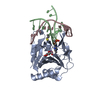
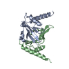
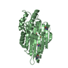
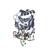
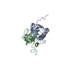
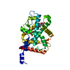
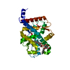
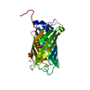
- PDB-5tg0: Crystal structure of the dimethylsulfoniopropionate (DMSP) lyase ... -

+
Open data


ID or keywords:

Loading...

-
Basic information

Entry
Database: PDB / ID: 5tg0
TitleCrystal structure of the dimethylsulfoniopropionate (DMSP) lyase DddK complexed with iron and zinc
Componentsdimethylsulfoniopropionate lyase DddK
KeywordsLYASE / DMSP lyase / Cupin / Nickel dependent / Metal binding
Function / homologyCupin 2, conserved barrel / Cupin domain / RmlC-like cupin domain superfamily / RmlC-like jelly roll fold / outer membrane-bounded periplasmic space / metal ion binding / : / Novel protein with potential Cupin domain
Function and homology information
Biological speciesPelagibacter ubique (bacteria)
MethodX-RAY DIFFRACTION / SYNCHROTRON / MOLECULAR REPLACEMENT / molecular replacement / Resolution: 1.44 Å
AuthorsSchnicker, N.J. / Dey, M.
Funding support United States, 1items
OrganizationGrant numberCountry
University of Iowa College of Liberal Arts and Sciences United States
CitationJournal: Biochemistry / Year: 2017
Title: Structural and Biochemical Insights into Dimethylsulfoniopropionate Cleavage by Cofactor-Bound DddK from the Prolific Marine Bacterium Pelagibacter.
Authors: Schnicker, N.J. / De Silva, S.M. / Todd, J.D. / Dey, M.
History
DepositionSep 27, 2016Deposition site: RCSB / Processing site: RCSB
Revision 1.0Jun 14, 2017Provider: repository / Type: Initial release
Revision 1.1Jun 21, 2017Group: Database references / Category: citation
Item: _citation.country / _citation.journal_id_ASTM ..._citation.country / _citation.journal_id_ASTM / _citation.journal_id_CSD / _citation.journal_volume / _citation.page_first / _citation.page_last
Revision 1.2Mar 6, 2024Group: Data collection / Database references / Derived calculations
Category: chem_comp_atom / chem_comp_bond ...chem_comp_atom / chem_comp_bond / database_2 / struct_conn
Item: _database_2.pdbx_DOI / _database_2.pdbx_database_accession ..._database_2.pdbx_DOI / _database_2.pdbx_database_accession / _struct_conn.pdbx_dist_value / _struct_conn.pdbx_ptnr1_label_alt_id / _struct_conn.ptnr1_label_atom_id / _struct_conn.ptnr2_auth_comp_id / _struct_conn.ptnr2_auth_seq_id / _struct_conn.ptnr2_label_asym_id / _struct_conn.ptnr2_label_atom_id / _struct_conn.ptnr2_label_comp_id

-
Structure visualization

Structure viewerMolecule:
MolmilJmol/JSmol

Downloads & links

-
Assembly

Deposited unit
A: dimethylsulfoniopropionate lyase DddK
hetero molecules


Theoretical massNumber of molelcules
Total (without water)16,7333
Polymers16,6121
Non-polymers1212
Water3,027168
1
A: dimethylsulfoniopropionate lyase DddK
hetero molecules

A: dimethylsulfoniopropionate lyase DddK
hetero molecules


Theoretical massNumber of molelcules
Total (without water)33,4666
Polymers33,2232
Non-polymers2434
Water362
TypeNameSymmetry operationNumber
identity operation1_555x,y,z1
crystal symmetry operation3_454-x-1,y,-z-1/21
Buried area3660 Å2
ΔGint-134 kcal/mol
Surface area13120 Å2
MethodPISA
Unit cell
Length a, b, c (Å)55.065, 85.416, 54.380
Angle α, β, γ (deg.)90.000, 90.000, 90.000
Int Tables number20
Space group name H-MC2221
Components on special symmetry positions
IDModelComponents
11A-454-

HOH

21A-460-

HOH

31A-468-

HOH

-
Components

#1: Protein dimethylsulfoniopropionate lyase DddK


Mass: 16611.645 Da / Num. of mol.: 1
Source method: isolated from a genetically manipulated source
Details: Residues 1-20 in the sequence are from the vector for the His tag.
Source: (gene. exp.) Pelagibacter ubique (strain HTCC1062) (bacteria)
Strain: HTCC1062 / Gene: SAR11_0394 / Production host: Escherichia coli (E. coli) / References: UniProt: Q4FNM4
#2: Chemical ChemComp-FE / FE (III) ION


Mass: 55.845 Da / Num. of mol.: 1 / Source method: obtained synthetically / Formula: Fe
#3: Chemical ChemComp-ZN / ZINC ION


Mass: 65.409 Da / Num. of mol.: 1 / Source method: obtained synthetically / Formula: Zn
#4: Water ChemComp-HOH / water


Mass: 18.015 Da / Num. of mol.: 168 / Source method: isolated from a natural source / Formula: H2O

-
Experimental details

-
Experiment

ExperimentMethod: X-RAY DIFFRACTION / Number of used crystals: 1

-
Sample preparation

CrystalDensity Matthews: 1.92 Å3/Da / Density % sol: 36.09 %
Crystal growTemperature: 293 K / Method: vapor diffusion, sitting drop / pH: 7.5 / Details: 0.1 M HEPES, 0.2 M NaCl, 20% PEG 3000

-
Data collection

DiffractionMean temperature: 100 K
Diffraction sourceSource: SYNCHROTRON / Site: APS / Beamline: 19-ID / Wavelength: 0.979 Å
DetectorType: ADSC QUANTUM 315 / Detector: CCD / Date: Nov 20, 2015
RadiationProtocol: SINGLE WAVELENGTH / Monochromatic (M) / Laue (L): M / Scattering type: x-ray
Radiation wavelengthWavelength: 0.979 Å / Relative weight: 1
ReflectionResolution: 1.44→46.28 Å / Num. obs: 23496 / % possible obs: 99.6 % / Redundancy: 6 % / Biso Wilson estimate: 12.65 Å2 / CC1/2: 0.999 / Rmerge(I) obs: 0.034 / Rpim(I) all: 0.015 / Rrim(I) all: 0.037 / Net I/σ(I): 29.8 / Num. measured all: 140675
Reflection shell
Resolution (Å)Redundancy (%)Rmerge(I) obsCC1/2Diffraction-ID% possible all
1.44-1.524.30.1690.971197.6
4.55-46.285.80.0250.999199.4

-
Phasing

PhasingMethod: molecular replacement

-
Processing

Software
NameVersionClassification
Aimless0.5.27data scaling
PHASERphasing
PHENIX1.10_2155refinement
PDB_EXTRACT3.2data extraction
XDSdata reduction
RefinementMethod to determine structure: MOLECULAR REPLACEMENT / Resolution: 1.44→27.532 Å / SU ML: 0.11 / Cross valid method: FREE R-VALUE / σ(F): 1.34 / Phase error: 12
RfactorNum. reflection% reflection
Rfree0.1399 2243 5.06 %
Rwork0.1101 --
obs0.1116 44346 98.8 %
Solvent computationShrinkage radii: 0.9 Å / VDW probe radii: 1.11 Å
Displacement parametersBiso max: 61.74 Å2 / Biso mean: 18.1367 Å2 / Biso min: 6.01 Å2
Refinement stepCycle: final / Resolution: 1.44→27.532 Å
ProteinNucleic acidLigandSolventTotal
Num. atoms1060 0 2 172 1234
Biso mean--9.79 31.02 -
Num. residues----137
Refine LS restraints
Refine-IDTypeDev idealNumber
X-RAY DIFFRACTIONf_bond_d0.011203
X-RAY DIFFRACTIONf_angle_d1.1051652
X-RAY DIFFRACTIONf_chiral_restr0.096176
X-RAY DIFFRACTIONf_plane_restr0.007217
X-RAY DIFFRACTIONf_dihedral_angle_d12.108439
LS refinement shell

Refine-ID: X-RAY DIFFRACTION / Total num. of bins used: 16

Resolution (Å)Rfactor RfreeNum. reflection RfreeRfactor RworkNum. reflection RworkNum. reflection all% reflection obs (%)
1.44-1.47130.20731190.152237235684
1.4713-1.50550.17081500.114626452795100
1.5055-1.54320.17661340.095827092843100
1.5432-1.58490.1751170.099326902807100
1.5849-1.63150.12821340.100426442778100
1.6315-1.68420.13971450.094826542799100
1.6842-1.74440.15651570.098726502807100
1.7444-1.81420.15831370.098226882825100
1.8142-1.89680.14371360.098426482784100
1.8968-1.99670.09441580.097326412799100
1.9967-2.12180.12881200.096426662786100
2.1218-2.28550.12561410.095126552796100
2.2855-2.51540.15841370.110926502787100
2.5154-2.87910.14511490.11992652280199
2.8791-3.6260.14111560.11832635279199
3.626-27.53750.13381530.12592639279299

+
About Yorodumi

-
News

-
Feb 9, 2022. New format data for meta-information of EMDB entries

New format data for meta-information of EMDB entries

  • Version 3 of the EMDB header file is now the official format.
  • The previous official version 1.9 will be removed from the archive.

Related info.:EMDB header

External links:wwPDB to switch to version 3 of the EMDB data model

-
Aug 12, 2020. Covid-19 info

Covid-19 info

URL: https://pdbjlvh1.pdbj.org/emnavi/covid19.php

New page: Covid-19 featured information page in EM Navigator.

Related info.:Covid-19 info / Mar 5, 2020. Novel coronavirus structure data

+
Mar 5, 2020. Novel coronavirus structure data

Novel coronavirus structure data

Related info.:Yorodumi Speices / Aug 12, 2020. Covid-19 info

External links:COVID-19 featured content - PDBj / Molecule of the Month (242):Coronavirus Proteases

+
Jan 31, 2019. EMDB accession codes are about to change! (news from PDBe EMDB page)

EMDB accession codes are about to change! (news from PDBe EMDB page)

  • The allocation of 4 digits for EMDB accession codes will soon come to an end. Whilst these codes will remain in use, new EMDB accession codes will include an additional digit and will expand incrementally as the available range of codes is exhausted. The current 4-digit format prefixed with “EMD-” (i.e. EMD-XXXX) will advance to a 5-digit format (i.e. EMD-XXXXX), and so on. It is currently estimated that the 4-digit codes will be depleted around Spring 2019, at which point the 5-digit format will come into force.
  • The EM Navigator/Yorodumi systems omit the EMD- prefix.

Related info.:Q: What is EMD? / ID/Accession-code notation in Yorodumi/EM Navigator

External links:EMDB Accession Codes are Changing Soon! / Contact to PDBj

+
Jul 12, 2017. Major update of PDB

Major update of PDB

  • wwPDB released updated PDB data conforming to the new PDBx/mmCIF dictionary.
  • This is a major update changing the version number from 4 to 5, and with Remediation, in which all the entries are updated.
  • In this update, many items about electron microscopy experimental information are reorganized (e.g. em_software).
  • Now, EM Navigator and Yorodumi are based on the updated data.

External links:wwPDB Remediation / Enriched Model Files Conforming to OneDep Data Standards Now Available in the PDB FTP Archive

-
Yorodumi

Thousand views of thousand structures

  • Yorodumi is a browser for structure data from EMDB, PDB, SASBDB, etc.
  • This page is also the successor to EM Navigator detail page, and also detail information page/front-end page for Omokage search.
  • The word "yorodu" (or yorozu) is an old Japanese word meaning "ten thousand". "mi" (miru) is to see.

Related info.:EMDB / PDB / SASBDB / Comparison of 3 databanks / Yorodumi Search / Aug 31, 2016. New EM Navigator & Yorodumi / Yorodumi Papers / Jmol/JSmol / Function and homology information / Changes in new EM Navigator and Yorodumi

Read more