 Movie
Movie Controller


+ Open data

- Basic information
| Entry | Database: PDB / ID: 6l1y | ||||||
|---|---|---|---|---|---|---|---|
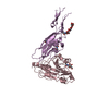
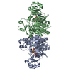
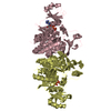
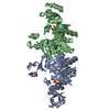
| Title | structure of gp120/CD4 with a non-canonical surface | ||||||
 Components Components |
| ||||||
 Keywords Keywords | PROTEIN BINDING / HIV / gp120 / CD4 receptor | ||||||
| Function / homology |  Function and homology information Function and homology informationhelper T cell enhancement of adaptive immune response / interleukin-16 binding / interleukin-16 receptor activity / maintenance of protein location in cell / cellular response to ionomycin / response to methamphetamine hydrochloride / T cell selection / MHC class II protein binding / interleukin-15-mediated signaling pathway / cellular response to granulocyte macrophage colony-stimulating factor stimulus ...helper T cell enhancement of adaptive immune response / interleukin-16 binding / interleukin-16 receptor activity / maintenance of protein location in cell / cellular response to ionomycin / response to methamphetamine hydrochloride / T cell selection / MHC class II protein binding / interleukin-15-mediated signaling pathway / cellular response to granulocyte macrophage colony-stimulating factor stimulus / positive regulation of monocyte differentiation / Alpha-defensins / Nef Mediated CD4 Down-regulation / regulation of T cell activation / response to vitamin D / Other interleukin signaling / extracellular matrix structural constituent / T cell receptor complex / enzyme-linked receptor protein signaling pathway / Translocation of ZAP-70 to Immunological synapse / Phosphorylation of CD3 and TCR zeta chains / Generation of second messenger molecules / macrophage differentiation / immunoglobulin binding / T cell differentiation / Co-inhibition by PD-1 / positive regulation of calcium ion transport into cytosol / Binding and entry of HIV virion / positive regulation of interleukin-2 production / coreceptor activity / positive regulation of T cell proliferation / positive regulation of calcium-mediated signaling / cell surface receptor protein tyrosine kinase signaling pathway / protein tyrosine kinase binding / host cell endosome membrane / clathrin-coated endocytic vesicle membrane / calcium-mediated signaling / Vpu mediated degradation of CD4 / MHC class II protein complex binding / response to estradiol / transmembrane signaling receptor activity / Downstream TCR signaling / Cargo recognition for clathrin-mediated endocytosis / T cell receptor signaling pathway / Clathrin-mediated endocytosis / virus receptor activity / signaling receptor activity / defense response to Gram-negative bacterium / adaptive immune response / response to ethanol / early endosome / cell surface receptor signaling pathway / cell adhesion / immune response / membrane raft / endoplasmic reticulum lumen / external side of plasma membrane / apoptotic process / viral envelope / symbiont entry into host cell / lipid binding / endoplasmic reticulum membrane / protein kinase binding / host cell plasma membrane / virion membrane / structural molecule activity / enzyme binding / signal transduction / protein homodimerization activity / zinc ion binding / identical protein binding / plasma membrane Similarity search - Function | ||||||
| Biological species |   Human immunodeficiency virus 1 Human immunodeficiency virus 1 Homo sapiens (human) Homo sapiens (human) | ||||||
| Method |  X-RAY DIFFRACTION / SYNCHROTRON / MOLECULAR REPLACEMENT / Resolution: 2.469 Å X-RAY DIFFRACTION / SYNCHROTRON / MOLECULAR REPLACEMENT / Resolution: 2.469 Å | ||||||
 Authors Authors | Liu, X. / Ning, W. | ||||||
| Funding support |  China, 1items China, 1items
| ||||||
 Citation Citation | Journal: Sci Rep / Year: 2017 Title: A non-canonical binding interface in the crystal structure of HIV-1 gp120 core in complex with CD4. Authors: Duan, L.W. / Zhang, H. / Zhao, M.T. / Sun, J.X. / Chen, W.L. / Lin, J.P. / Liu, X.Q. | ||||||
| History |
|
- Structure visualization
| Structure viewer | Molecule: MolmilJmol/JSmol |
|---|
- Downloads & links
-Download
| PDBx/mmCIF format | 6l1y.cif.gz | 123.3 KB | Display | PDBx/mmCIF format |
|---|---|---|---|---|
| PDB format | pdb6l1y.ent.gz | 92 KB | Display | PDB format |
| PDBx/mmJSON format | 6l1y.json.gz | Tree view | PDBx/mmJSON format | |
| Others |  Other downloads Other downloads |
-Validation report
| Arichive directory | https://data.pdbj.org/pub/pdb/validation_reports/l1/6l1yftp://data.pdbj.org/pub/pdb/validation_reports/l1/6l1y | HTTPS FTP |
|---|
-Related structure data
| Related structure data |  3tgtS S: Starting model for refinement |
|---|---|
| Similar structure data |


-Links
 PDBj
PDBj


- Assembly
| Deposited unit | 
| ||||||||
|---|---|---|---|---|---|---|---|---|---|
| 1 |
| ||||||||
| Unit cell |
|
-Components
| #1: Protein | Mass: 43340.457 Da / Num. of mol.: 1 Source method: isolated from a genetically manipulated source Source: (gene. exp.) Human immunodeficiency virus 1Production host: Insect expression vector pBlueBacmsGCA1His (others) References: UniProt: G4XFJ5*PLUS | ||||||||
|---|---|---|---|---|---|---|---|---|---|
| #2: Protein | Mass: 19654.334 Da / Num. of mol.: 1 Source method: isolated from a genetically manipulated source Source: (gene. exp.) Homo sapiens (human) / Gene: CD4Production host: Insect expression vector pBlueBacmsGCA1His (others) References: UniProt: P01730 | ||||||||
| #3: Sugar | ChemComp-NAG /   Type: D-saccharide, beta linking / Mass: 221.208 Da / Num. of mol.: 8 Type: D-saccharide, beta linking / Mass: 221.208 Da / Num. of mol.: 8Source method: isolated from a genetically manipulated source Formula: C8H15NO6 / Feature type: SUBJECT OF INVESTIGATION #4: Chemical | ChemComp-EPE / |   Mass: 238.305 Da / Num. of mol.: 1 / Source method: obtained synthetically / Formula: C8H18N2O4S / Comment: pH buffer*YM Mass: 238.305 Da / Num. of mol.: 1 / Source method: obtained synthetically / Formula: C8H18N2O4S / Comment: pH buffer*YM#5: Water | ChemComp-HOH / |  Mass: 18.015 Da / Num. of mol.: 123 / Source method: isolated from a natural source / Formula: H2O Mass: 18.015 Da / Num. of mol.: 123 / Source method: isolated from a natural source / Formula: H2OHas ligand of interest | Y | Has protein modification | Y | |
-Experimental details
-Experiment
| Experiment | Method: X-RAY DIFFRACTION / Number of used crystals: 1 |
|---|
- Sample preparation
| Crystal | Density Matthews: 2.78 Å3/Da / Density % sol: 55.68 % |
|---|---|
| Crystal grow | Temperature: 293 K / Method: evaporation / pH: 7.4 Details: 0.1 M HEPES sodium, pH 7.4, 11% PEG 4000 and 10% 2-propanol |
-Data collection
| Diffraction | Mean temperature: 100 K / Serial crystal experiment: N |
|---|---|
| Diffraction source | Source: SYNCHROTRON / Site: BSRF / Beamline: 3W1A / Wavelength: 0.9792 Å |
| Detector | Type: MARMOSAIC 225 mm CCD / Detector: CCD / Date: Oct 25, 2013 |
| Radiation | Protocol: SINGLE WAVELENGTH / Monochromatic (M) / Laue (L): M / Scattering type: x-ray |
| Radiation wavelength | Wavelength: 0.9792 Å / Relative weight: 1 |
| Reflection | Resolution: 2.469→50 Å / Num. obs: 21690 / % possible obs: 95.6 % / Redundancy: 3 % / Biso Wilson estimate: 49.6 Å2 / Rmerge(I) obs: 0.05 / Rsym value: 0.03 / Net I/σ(I): 21.6 |
| Reflection shell | Resolution: 2.47→2.49 Å / Redundancy: 2.7 % / Rmerge(I) obs: 0.47 / Mean I/σ(I) obs: 2.15 / Num. unique obs: 1803 / Rsym value: 0.32 / % possible all: 96.8 |
- Processing
| Software |
| ||||||||||||||||||||||||||||||||||||||||||||||||||||||
|---|---|---|---|---|---|---|---|---|---|---|---|---|---|---|---|---|---|---|---|---|---|---|---|---|---|---|---|---|---|---|---|---|---|---|---|---|---|---|---|---|---|---|---|---|---|---|---|---|---|---|---|---|---|---|---|
| Refinement | Method to determine structure: MOLECULAR REPLACEMENT Starting model: 3TGT Resolution: 2.469→33.815 Å / SU ML: 0.37 / Cross valid method: THROUGHOUT / σ(F): 1.35 / Phase error: 30.01
| ||||||||||||||||||||||||||||||||||||||||||||||||||||||
| Solvent computation | Shrinkage radii: 0.9 Å / VDW probe radii: 1.11 Å | ||||||||||||||||||||||||||||||||||||||||||||||||||||||
| Displacement parameters | Biso max: 117.59 Å2 / Biso mean: 49.87 Å2 / Biso min: 22.58 Å2 | ||||||||||||||||||||||||||||||||||||||||||||||||||||||
| Refinement step | Cycle: final / Resolution: 2.469→33.815 Å
| ||||||||||||||||||||||||||||||||||||||||||||||||||||||
| Refine LS restraints |
| ||||||||||||||||||||||||||||||||||||||||||||||||||||||
| LS refinement shell | Refine-ID: X-RAY DIFFRACTION / Rfactor Rfree error: 0
|
