 Movie
Movie Controller

[English] 日本語
 Yorodumi
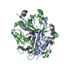
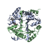
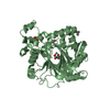
- PDB-6l0u: Crystal structure of mouse glyoxalase I complexed with a small mo... -
+ Open data

- Basic information
| Entry | Database: PDB / ID: 6l0u | ||||||
|---|---|---|---|---|---|---|---|
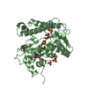
| Title | Crystal structure of mouse glyoxalase I complexed with a small molecule inhibitor | ||||||
 Components Components | Lactoylglutathione lyase | ||||||
 Keywords Keywords | LYASE/LYASE INHIBITOR / mouse glyoxalase I / inhibitor / LYASE / LYASE-LYASE INHIBITOR complex | ||||||
| Function / homology |  Function and homology information Function and homology informationPyruvate metabolism / lactoylglutathione lyase / lactoylglutathione lyase activity / : / methylglyoxal metabolic process / osteoclast differentiation / glutathione metabolic process / regulation of transcription by RNA polymerase II / negative regulation of apoptotic process / zinc ion binding ...Pyruvate metabolism / lactoylglutathione lyase / lactoylglutathione lyase activity / : / methylglyoxal metabolic process / osteoclast differentiation / glutathione metabolic process / regulation of transcription by RNA polymerase II / negative regulation of apoptotic process / zinc ion binding / nucleoplasm / plasma membrane / cytosol Similarity search - Function | ||||||
| Biological species |  | ||||||
| Method |  X-RAY DIFFRACTION / SYNCHROTRON / MOLECULAR REPLACEMENT / Resolution: 1.95 Å X-RAY DIFFRACTION / SYNCHROTRON / MOLECULAR REPLACEMENT / Resolution: 1.95 Å | ||||||
 Authors Authors | Jiang, L.L. / Zhou, L. | ||||||
 Citation Citation | Journal: To Be Published Title: Crystal structure of mouse glyoxalase I complexed with a small molecule inhibitor Authors: Jiang, L.L. / Zhou, L. | ||||||
| History |
|
- Structure visualization
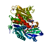
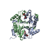
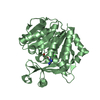
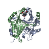
| Structure viewer | Molecule: MolmilJmol/JSmol |
|---|
- Downloads & links
-Download
| PDBx/mmCIF format | 6l0u.cif.gz | 89.5 KB | Display | PDBx/mmCIF format |
|---|---|---|---|---|
| PDB format | pdb6l0u.ent.gz | 65.4 KB | Display | PDB format |
| PDBx/mmJSON format | 6l0u.json.gz | Tree view | PDBx/mmJSON format | |
| Others |  Other downloads Other downloads |
-Validation report
| Arichive directory | https://data.pdbj.org/pub/pdb/validation_reports/l0/6l0uftp://data.pdbj.org/pub/pdb/validation_reports/l0/6l0u | HTTPS FTP |
|---|
-Related structure data
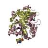
| Related structure data |  2za0S S: Starting model for refinement |
|---|---|
| Similar structure data |

-Links
 PDBj
PDBj- Assembly
| Deposited unit | 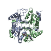
| |||||||||||||||||||||||||||||||||
|---|---|---|---|---|---|---|---|---|---|---|---|---|---|---|---|---|---|---|---|---|---|---|---|---|---|---|---|---|---|---|---|---|---|---|
| 1 |
| |||||||||||||||||||||||||||||||||
| Unit cell |
| |||||||||||||||||||||||||||||||||
| Noncrystallographic symmetry (NCS) | NCS domain:
NCS domain segments: Component-ID: 1 / Ens-ID: 1 / End auth comp-ID: ILE / End label comp-ID: ILE
|
-Components
| #1: Protein | Mass: 20278.953 Da / Num. of mol.: 2 Source method: isolated from a genetically manipulated source Source: (gene. exp.)  #2: Chemical |   Mass: 65.409 Da / Num. of mol.: 2 / Source method: obtained synthetically / Formula: Zn Mass: 65.409 Da / Num. of mol.: 2 / Source method: obtained synthetically / Formula: Zn#3: Chemical | ChemComp-E1L / | 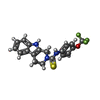  Mass: 391.410 Da / Num. of mol.: 1 / Source method: obtained synthetically / Formula: C19H16F3N3OS / Feature type: SUBJECT OF INVESTIGATION Mass: 391.410 Da / Num. of mol.: 1 / Source method: obtained synthetically / Formula: C19H16F3N3OS / Feature type: SUBJECT OF INVESTIGATION#4: Water | ChemComp-HOH / |  Mass: 18.015 Da / Num. of mol.: 108 / Source method: isolated from a natural source / Formula: H2O Mass: 18.015 Da / Num. of mol.: 108 / Source method: isolated from a natural source / Formula: H2OHas ligand of interest | Y | |
|---|
-Experimental details
-Experiment
| Experiment | Method: X-RAY DIFFRACTION / Number of used crystals: 1 |
|---|
- Sample preparation
| Crystal | Density Matthews: 2.15 Å3/Da / Density % sol: 42.92 % |
|---|---|
| Crystal grow | Temperature: 289 K / Method: vapor diffusion, hanging drop / pH: 6 Details: 0.1 M MES pH 6.0, 12% (w/v) PEG2000, 16% v/v 2-Propanol |
-Data collection
| Diffraction | Mean temperature: 100 K / Serial crystal experiment: N | ||||||||||||||||||||||||||||||||||||||||||||||||||||||||||||||||||||||||||||||||||||||||
|---|---|---|---|---|---|---|---|---|---|---|---|---|---|---|---|---|---|---|---|---|---|---|---|---|---|---|---|---|---|---|---|---|---|---|---|---|---|---|---|---|---|---|---|---|---|---|---|---|---|---|---|---|---|---|---|---|---|---|---|---|---|---|---|---|---|---|---|---|---|---|---|---|---|---|---|---|---|---|---|---|---|---|---|---|---|---|---|---|---|
| Diffraction source | Source: SYNCHROTRON / Site: SSRF  / Beamline: BL18U1 / Wavelength: 0.97853 Å / Beamline: BL18U1 / Wavelength: 0.97853 Å | ||||||||||||||||||||||||||||||||||||||||||||||||||||||||||||||||||||||||||||||||||||||||
| Detector | Type: ADSC QUANTUM 315r / Detector: CCD / Date: May 10, 2019 | ||||||||||||||||||||||||||||||||||||||||||||||||||||||||||||||||||||||||||||||||||||||||
| Radiation | Protocol: SINGLE WAVELENGTH / Monochromatic (M) / Laue (L): M / Scattering type: x-ray | ||||||||||||||||||||||||||||||||||||||||||||||||||||||||||||||||||||||||||||||||||||||||
| Radiation wavelength | Wavelength: 0.97853 Å / Relative weight: 1 | ||||||||||||||||||||||||||||||||||||||||||||||||||||||||||||||||||||||||||||||||||||||||
| Reflection | Resolution: 1.95→50 Å / Num. obs: 25098 / % possible obs: 100 % / Redundancy: 6.7 % / Rmerge(I) obs: 0.067 / Rpim(I) all: 0.028 / Rrim(I) all: 0.073 / Χ2: 0.663 / Net I/σ(I): 5.3 / Num. measured all: 167399 | ||||||||||||||||||||||||||||||||||||||||||||||||||||||||||||||||||||||||||||||||||||||||
| Reflection shell | Diffraction-ID: 1 / % possible all: 100
|
- Processing
| Software |
| ||||||||||||||||||||||||||||||||||||||||||||||||||||||||||||
|---|---|---|---|---|---|---|---|---|---|---|---|---|---|---|---|---|---|---|---|---|---|---|---|---|---|---|---|---|---|---|---|---|---|---|---|---|---|---|---|---|---|---|---|---|---|---|---|---|---|---|---|---|---|---|---|---|---|---|---|---|---|
| Refinement | Method to determine structure: MOLECULAR REPLACEMENT Starting model: 2ZA0 Resolution: 1.95→23.16 Å / SU ML: 0.22 / Cross valid method: THROUGHOUT / σ(F): 1.38 / Phase error: 25.46
| ||||||||||||||||||||||||||||||||||||||||||||||||||||||||||||
| Solvent computation | Shrinkage radii: 0.9 Å / VDW probe radii: 1.11 Å | ||||||||||||||||||||||||||||||||||||||||||||||||||||||||||||
| Displacement parameters | Biso max: 88.82 Å2 / Biso mean: 41.79 Å2 / Biso min: 18.66 Å2 | ||||||||||||||||||||||||||||||||||||||||||||||||||||||||||||
| Refinement step | Cycle: final / Resolution: 1.95→23.16 Å
| ||||||||||||||||||||||||||||||||||||||||||||||||||||||||||||
| Refine LS restraints |
| ||||||||||||||||||||||||||||||||||||||||||||||||||||||||||||
| Refine LS restraints NCS |
| ||||||||||||||||||||||||||||||||||||||||||||||||||||||||||||
| LS refinement shell | Refine-ID: X-RAY DIFFRACTION / Rfactor Rfree error: 0
|
