 ムービー
ムービー コントローラー


+ データを開く

- 基本情報
基本情報
| 登録情報 | データベース: PDB / ID: 6kto | ||||||
|---|---|---|---|---|---|---|---|
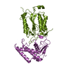
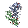
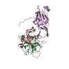
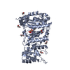
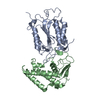
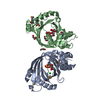
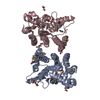
| タイトル | Crystal structure of human SHLD3-C-REV7-O-REV7-SHLD2 complex | ||||||
 要素 要素 |
| ||||||
 キーワード キーワード | REPLICATION / Shieldin / Complex / Conformational dimer / NHEJ | ||||||
| 機能・相同性 |  機能・相同性情報 機能・相同性情報somatic diversification of immunoglobulins involved in immune response / DNA damage response, signal transduction resulting in transcription / telomere maintenance in response to DNA damage / negative regulation of transcription regulatory region DNA binding / zeta DNA polymerase complex / positive regulation of isotype switching / positive regulation of extracellular matrix assembly / negative regulation of transcription by competitive promoter binding / negative regulation of cell-cell adhesion mediated by cadherin / JUN kinase binding ...somatic diversification of immunoglobulins involved in immune response / DNA damage response, signal transduction resulting in transcription / telomere maintenance in response to DNA damage / negative regulation of transcription regulatory region DNA binding / zeta DNA polymerase complex / positive regulation of isotype switching / positive regulation of extracellular matrix assembly / negative regulation of transcription by competitive promoter binding / negative regulation of cell-cell adhesion mediated by cadherin / JUN kinase binding / negative regulation of epithelial to mesenchymal transition / negative regulation of ubiquitin protein ligase activity / regulation of double-strand break repair via homologous recombination / mitotic spindle assembly checkpoint signaling / positive regulation of double-strand break repair via nonhomologous end joining / negative regulation of double-strand break repair via homologous recombination / error-prone translesion synthesis / positive regulation of epithelial to mesenchymal transition / Translesion synthesis by REV1 / Translesion synthesis by POLK / Translesion synthesis by POLI / regulation of cell growth / actin filament organization / negative regulation of canonical Wnt signaling pathway / negative regulation of protein catabolic process / negative regulation of DNA-binding transcription factor activity / spindle / double-strand break repair / actin cytoskeleton / positive regulation of peptidyl-serine phosphorylation / chromosome / site of double-strand break / RNA polymerase II-specific DNA-binding transcription factor binding / cell division / DNA repair / chromatin / nucleolus / positive regulation of DNA-templated transcription / negative regulation of transcription by RNA polymerase II / nucleoplasm / nucleus / cytoplasm 類似検索 - 分子機能 | ||||||
| 生物種 |  Homo sapiens (ヒト) Homo sapiens (ヒト) | ||||||
| 手法 |  X線回折 / シンクロトロン / 分子置換 / 解像度: 3.44976331487 Å X線回折 / シンクロトロン / 分子置換 / 解像度: 3.44976331487 Å | ||||||
 データ登録者 データ登録者 | Liang, L. / Yin, Y. | ||||||
| 資金援助 |  中国, 1件 中国, 1件
| ||||||
 引用 引用 | ジャーナル: Nat Commun / 年: 2020 タイトル: Molecular basis for assembly of the shieldin complex and its implications for NHEJ. 著者: Liang, L. / Feng, J. / Zuo, P. / Yang, J. / Lu, Y. / Yin, Y. | ||||||
| 履歴 |
|
- 構造の表示
構造の表示
| 構造ビューア | 分子: MolmilJmol/JSmol |
|---|
- ダウンロードとリンク
-ダウンロード
| PDBx/mmCIF形式 | 6kto.cif.gz | 114.2 KB | 表示 | PDBx/mmCIF形式 |
|---|---|---|---|---|
| PDB形式 | pdb6kto.ent.gz | 80.7 KB | 表示 | PDB形式 |
| PDBx/mmJSON形式 | 6kto.json.gz | ツリー表示 | PDBx/mmJSON形式 | |
| その他 |  その他のダウンロード その他のダウンロード |
-検証レポート
| 文書・要旨 | 6kto_validation.pdf.gz | 470.4 KB | 表示 | wwPDB検証レポート |
|---|---|---|---|---|
| 文書・詳細版 | 6kto_full_validation.pdf.gz | 491 KB | 表示 | |
| XML形式データ | 6kto_validation.xml.gz | 20.3 KB | 表示 | |
| CIF形式データ | 6kto_validation.cif.gz | 26.7 KB | 表示 | |
| アーカイブディレクトリ | https://data.pdbj.org/pub/pdb/validation_reports/kt/6ktoftp://data.pdbj.org/pub/pdb/validation_reports/kt/6kto | HTTPS FTP |
-関連構造データ
| 関連構造データ | 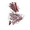 3vu7S S: 精密化の開始モデル |
|---|---|
| 類似構造データ |


-リンク
 PDBj
PDBj


- 集合体
集合体
| 登録構造単位 | 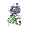
| ||||||||||||
|---|---|---|---|---|---|---|---|---|---|---|---|---|---|
| 1 |
| ||||||||||||
| 単位格子 |
|
-要素
| #1: タンパク質 | 分子量: 26187.352 Da / 分子数: 2 / 由来タイプ: 組換発現 / 由来: (組換発現) Homo sapiens (ヒト) / 遺伝子: MAD2L2, MAD2B, REV7 / 発現宿主:  #2: タンパク質 | | 分子量: 7402.546 Da / 分子数: 1 / 由来タイプ: 組換発現 / 由来: (組換発現) Homo sapiens (ヒト) / 遺伝子: SHLD3, FLJ26957, RINN1 / 発現宿主: #3: タンパク質 | | 分子量: 5953.904 Da / 分子数: 1 / 由来タイプ: 組換発現 / 由来: (組換発現) Homo sapiens (ヒト) / 遺伝子: SHLD2, FAM35A, RINN2 / 発現宿主: |
|---|
-実験情報
-実験
| 実験 | 手法: X線回折 / 使用した結晶の数: 1 |
|---|
- 試料調製
試料調製
| 結晶 | マシュー密度: 3.15 Å3/Da / 溶媒含有率: 60.9 % |
|---|---|
| 結晶化 | 温度: 293 K / 手法: 蒸気拡散法, シッティングドロップ法 詳細: 0.02M Magnesium chloride hexahydrate, 0.1M HEPES 7.5, 22% w/v Poly (acrylic acid sodium salt) 5100 |
-データ収集
| 回折 | 平均測定温度: 100 K / Serial crystal experiment: N |
|---|---|
| 放射光源 | 由来: シンクロトロン / サイト: SSRF / ビームライン: BL18U1 / 波長: 0.97853 Å |
| 検出器 | タイプ: DECTRIS PILATUS 6M / 検出器: PIXEL / 日付: 2019年1月5日 |
| 放射 | プロトコル: SINGLE WAVELENGTH / 単色(M)・ラウエ(L): M / 散乱光タイプ: x-ray |
| 放射波長 | 波長: 0.97853 Å / 相対比: 1 |
| 反射 | 解像度: 3.44→50 Å / Num. obs: 11237 / % possible obs: 100 % / 冗長度: 13 % / Biso Wilson estimate: 58.7354852027 Å2 / Rpim(I) all: 0.058 / Net I/σ(I): 12.75 |
| 反射 シェル | 解像度: 3.45→3.72 Å / Num. unique obs: 2312 / Rpim(I) all: 0.452 |
- 解析
解析
| ソフトウェア |
| |||||||||||||||||||||||||||||||||||
|---|---|---|---|---|---|---|---|---|---|---|---|---|---|---|---|---|---|---|---|---|---|---|---|---|---|---|---|---|---|---|---|---|---|---|---|---|
| 精密化 | 構造決定の手法: 分子置換 開始モデル: 3vu7 解像度: 3.44976331487→46.4411009446 Å / SU ML: 0.408163546905 / 交差検証法: FREE R-VALUE / σ(F): 1.36702633773 / 位相誤差: 24.9565654608 立体化学のターゲット値: GeoStd + Monomer Library + CDL v1.2
| |||||||||||||||||||||||||||||||||||
| 溶媒の処理 | 減衰半径: 0.9 Å / VDWプローブ半径: 1.11 Å / 溶媒モデル: FLAT BULK SOLVENT MODEL | |||||||||||||||||||||||||||||||||||
| 原子変位パラメータ | Biso mean: 47.6481043069 Å2 | |||||||||||||||||||||||||||||||||||
| 精密化ステップ | サイクル: LAST / 解像度: 3.44976331487→46.4411009446 Å
| |||||||||||||||||||||||||||||||||||
| 拘束条件 |
| |||||||||||||||||||||||||||||||||||
| LS精密化 シェル |
|
