[English] 日本語
 Yorodumi
- PDB-6krn: Crystal structure of GH30 xylanase B from Talaromyces cellulolyti... -
+ Open data
- Basic information
| Entry | Database: PDB / ID: 6krn | |||||||||
|---|---|---|---|---|---|---|---|---|---|---|
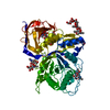
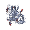
| Title | Crystal structure of GH30 xylanase B from Talaromyces cellulolyticus expressed by Pichia pastoris in complex with aldotriuronic acid | |||||||||
 Components Components | Mating factor alpha,GH30 Xylanase B | |||||||||
 Keywords Keywords | HYDROLASE / Xylanase / Glucuronoxylanase | |||||||||
| Function / homology |  Function and homology information Function and homology informationmating pheromone activity / mating / pheromone-dependent signal transduction involved in conjugation with cellular fusion / glucosylceramide catabolic process / glucosylceramidase activity / xylan catabolic process / extracellular region / membrane Similarity search - Function | |||||||||
| Biological species |   Talaromyces cellulolyticus CF-2612 (fungus) Talaromyces cellulolyticus CF-2612 (fungus) | |||||||||
| Method |  X-RAY DIFFRACTION / X-RAY DIFFRACTION /  SYNCHROTRON / SYNCHROTRON /  MOLECULAR REPLACEMENT / Resolution: 1.653 Å MOLECULAR REPLACEMENT / Resolution: 1.653 Å | |||||||||
 Authors Authors | Nakamichi, Y. / Watanabe, M. / Inoue, H. | |||||||||
 Citation Citation |  Journal: Febs Open Bio / Year: 2020 Journal: Febs Open Bio / Year: 2020Title: Substrate recognition by a bifunctional GH30-7 xylanase B from Talaromyces cellulolyticus. Authors: Nakamichi, Y. / Watanabe, M. / Matsushika, A. / Inoue, H. | |||||||||
| History |
|
- Structure visualization
| Structure viewer | Molecule:  Molmil Molmil Jmol/JSmol Jmol/JSmol |
|---|
- Downloads & links
- Download
| PDBx/mmCIF format |  6krn.cif.gz 6krn.cif.gz | 128.6 KB | Display |  PDBx/mmCIF format PDBx/mmCIF format |
|---|---|---|---|---|
| PDB format |  pdb6krn.ent.gz pdb6krn.ent.gz | 93.4 KB | Display |  PDB format PDB format |
| PDBx/mmJSON format |  6krn.json.gz 6krn.json.gz | Tree view |  PDBx/mmJSON format PDBx/mmJSON format | |
| Others |  Other downloads Other downloads |
-Validation report
| Arichive directory |  https://data.pdbj.org/pub/pdb/validation_reports/kr/6krn https://data.pdbj.org/pub/pdb/validation_reports/kr/6krn ftp://data.pdbj.org/pub/pdb/validation_reports/kr/6krn ftp://data.pdbj.org/pub/pdb/validation_reports/kr/6krn | HTTPS FTP |
|---|
-Related structure data
| Related structure data |  6krlC  6iujS C: citing same article ( S: Starting model for refinement |
|---|---|
| Similar structure data |
- Links
Links
- Assembly
| Deposited unit | 
| ||||||||
|---|---|---|---|---|---|---|---|---|---|
| 1 |
| ||||||||
| Unit cell |
|
- Components
-Protein / Non-polymers , 2 types, 499 molecules A

| #1: Protein | Mass: 59301.277 Da / Num. of mol.: 1 Source method: isolated from a genetically manipulated source Details: The fusion protein of Mating factor alpha (UNP residues 1-89), linker, and GH30 Xylanase B (UNP residues 23-474). Source: (gene. exp.)   Talaromyces cellulolyticus CF-2612 (fungus) Talaromyces cellulolyticus CF-2612 (fungus)Production host:  Komagataella phaffii GS115 (fungus) Komagataella phaffii GS115 (fungus)References: UniProt: P25501, UniProt: A0A4V8H018, UniProt: A0A510NXC4*PLUS |
|---|---|
| #6: Water | ChemComp-HOH / |
-Sugars , 4 types, 6 molecules
| #2: Polysaccharide | Source method: isolated from a genetically manipulated source #3: Polysaccharide | Source method: isolated from a genetically manipulated source #4: Polysaccharide | 4-O-methyl-alpha-D-glucopyranuronic acid-(1-2)-beta-D-xylopyranose-(1-4)-beta-D-xylopyranose | Source method: isolated from a genetically manipulated source #5: Polysaccharide | beta-D-mannopyranose-(1-4)-2-acetamido-2-deoxy-beta-D-glucopyranose-(1-4)-2-acetamido-2-deoxy-beta- ...beta-D-mannopyranose-(1-4)-2-acetamido-2-deoxy-beta-D-glucopyranose-(1-4)-2-acetamido-2-deoxy-beta-D-glucopyranose | Source method: isolated from a genetically manipulated source |
|---|
-Details
| Has ligand of interest | Y |
|---|---|
| Has protein modification | Y |
| Sequence details | The sequence region is from a commercial vector pPIC9K (Thermo fisher scientific). |
-Experimental details
-Experiment
| Experiment | Method:  X-RAY DIFFRACTION / Number of used crystals: 1 X-RAY DIFFRACTION / Number of used crystals: 1 |
|---|
- Sample preparation
| Crystal | Density Matthews: 2.49 Å3/Da / Density % sol: 50.5 % |
|---|---|
| Crystal grow | Temperature: 293 K / Method: vapor diffusion, hanging drop / pH: 7.3 / Details: PEG 3350, HEPES, magnesium chloride |
-Data collection
| Diffraction | Mean temperature: 173 K / Serial crystal experiment: N |
|---|---|
| Diffraction source | Source:  SYNCHROTRON / Site: SYNCHROTRON / Site:  SPring-8 SPring-8  / Beamline: BL44XU / Wavelength: 0.9 Å / Beamline: BL44XU / Wavelength: 0.9 Å |
| Detector | Type: DECTRIS EIGER X 16M / Detector: PIXEL / Date: Jun 15, 2019 |
| Radiation | Protocol: SINGLE WAVELENGTH / Monochromatic (M) / Laue (L): M / Scattering type: x-ray |
| Radiation wavelength | Wavelength: 0.9 Å / Relative weight: 1 |
| Reflection | Resolution: 1.653→33.41 Å / Num. obs: 71087 / % possible obs: 99.41 % / Redundancy: 4.5 % / Biso Wilson estimate: 22.31 Å2 / CC1/2: 0.999 / Rmerge(I) obs: 0.04648 / Rpim(I) all: 0.02424 / Rrim(I) all: 0.05262 / Net I/σ(I): 15.35 |
| Reflection shell | Resolution: 1.653→1.712 Å / Redundancy: 4.5 % / Rmerge(I) obs: 0.458 / Mean I/σ(I) obs: 2.34 / Num. unique obs: 7017 / CC1/2: 0.847 / Rpim(I) all: 0.2389 / Rrim(I) all: 0.5184 / % possible all: 99.03 |
- Processing
| Software |
| ||||||||||||||||||||||||||||||||||||||||||||||||||||||||||||||||||||||||||||||||||||||||||||||||||||||||||||||||||||||||||||||||||||||||||||||||||||||
|---|---|---|---|---|---|---|---|---|---|---|---|---|---|---|---|---|---|---|---|---|---|---|---|---|---|---|---|---|---|---|---|---|---|---|---|---|---|---|---|---|---|---|---|---|---|---|---|---|---|---|---|---|---|---|---|---|---|---|---|---|---|---|---|---|---|---|---|---|---|---|---|---|---|---|---|---|---|---|---|---|---|---|---|---|---|---|---|---|---|---|---|---|---|---|---|---|---|---|---|---|---|---|---|---|---|---|---|---|---|---|---|---|---|---|---|---|---|---|---|---|---|---|---|---|---|---|---|---|---|---|---|---|---|---|---|---|---|---|---|---|---|---|---|---|---|---|---|---|---|---|---|
| Refinement | Method to determine structure:  MOLECULAR REPLACEMENT MOLECULAR REPLACEMENTStarting model: 6IUJ Resolution: 1.653→33.41 Å / Cor.coef. Fo:Fc: 0.967 / Cor.coef. Fo:Fc free: 0.962 / Cross valid method: FREE R-VALUE / ESU R: 0.084 / ESU R Free: 0.081 Details: Hydrogens have been added in their riding positions
| ||||||||||||||||||||||||||||||||||||||||||||||||||||||||||||||||||||||||||||||||||||||||||||||||||||||||||||||||||||||||||||||||||||||||||||||||||||||
| Solvent computation | Ion probe radii: 0.8 Å / Shrinkage radii: 0.8 Å / VDW probe radii: 1.2 Å | ||||||||||||||||||||||||||||||||||||||||||||||||||||||||||||||||||||||||||||||||||||||||||||||||||||||||||||||||||||||||||||||||||||||||||||||||||||||
| Displacement parameters | Biso mean: 23.318 Å2
| ||||||||||||||||||||||||||||||||||||||||||||||||||||||||||||||||||||||||||||||||||||||||||||||||||||||||||||||||||||||||||||||||||||||||||||||||||||||
| Refinement step | Cycle: LAST / Resolution: 1.653→33.41 Å
| ||||||||||||||||||||||||||||||||||||||||||||||||||||||||||||||||||||||||||||||||||||||||||||||||||||||||||||||||||||||||||||||||||||||||||||||||||||||
| Refine LS restraints |
| ||||||||||||||||||||||||||||||||||||||||||||||||||||||||||||||||||||||||||||||||||||||||||||||||||||||||||||||||||||||||||||||||||||||||||||||||||||||
| LS refinement shell |
|
 Movie
Movie Controller


 PDBj
PDBj