 Movie
Movie Controller

[English] 日本語
 Yorodumi
- PDB-6kkw: Crystal structure of the complex of phosphopantetheine adenylyl t... -
+ Open data

- Basic information
| Entry | Database: PDB / ID: 6kkw | ||||||
|---|---|---|---|---|---|---|---|
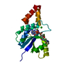
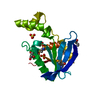
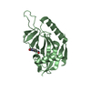
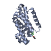
| Title | Crystal structure of the complex of phosphopantetheine adenylyl transferase from Acinetobacter baumannii with Dephospho Coenzyme at 3.2 A resolution. | ||||||
 Components Components | Phosphopantetheine adenylyltransferase | ||||||
 Keywords Keywords | TRANSFERASE | ||||||
| Function / homology |  Function and homology information Function and homology informationpantetheine-phosphate adenylyltransferase / pantetheine-phosphate adenylyltransferase activity / coenzyme A biosynthetic process / ATP binding / cytoplasm Similarity search - Function | ||||||
| Biological species |  Acinetobacter baumannii (bacteria) Acinetobacter baumannii (bacteria) | ||||||
| Method |  X-RAY DIFFRACTION / SYNCHROTRON / MOLECULAR REPLACEMENT / Resolution: 3.202 Å X-RAY DIFFRACTION / SYNCHROTRON / MOLECULAR REPLACEMENT / Resolution: 3.202 Å | ||||||
 Authors Authors | Viswanathan, V. / Gupta, A. / Sharma, S. / Singh, T.P. | ||||||
 Citation Citation | Journal: To Be Published Title: Crystal structure of the complex of phosphopantetheine adenylyl transferase from Acinetobacter baumannii with Dephospho Coenzyme at 3.2 A resolution. Authors: Viswanathan, V. / Gupta, A. / Sharma, S. / Singh, T.P. | ||||||
| History |
|
- Structure visualization
| Structure viewer | Molecule: MolmilJmol/JSmol |
|---|
- Downloads & links
-Download
| PDBx/mmCIF format | 6kkw.cif.gz | 51.3 KB | Display | PDBx/mmCIF format |
|---|---|---|---|---|
| PDB format | pdb6kkw.ent.gz | 35.2 KB | Display | PDB format |
| PDBx/mmJSON format | 6kkw.json.gz | Tree view | PDBx/mmJSON format | |
| Others |  Other downloads Other downloads |
-Validation report
| Arichive directory | https://data.pdbj.org/pub/pdb/validation_reports/kk/6kkwftp://data.pdbj.org/pub/pdb/validation_reports/kk/6kkw | HTTPS FTP |
|---|
-Related structure data
| Related structure data | 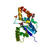 6jogS S: Starting model for refinement |
|---|---|
| Similar structure data |


-Links
 PDBj
PDBj- Assembly
| Deposited unit | 
| ||||||||
|---|---|---|---|---|---|---|---|---|---|
| 1 |
| ||||||||
| Unit cell |
|
-Components
| #1: Protein | Mass: 18479.033 Da / Num. of mol.: 1 Source method: isolated from a genetically manipulated source Source: (gene. exp.) Acinetobacter baumannii (bacteria) / Gene: coaD / Production host: References: UniProt: A0A059ZFC5, UniProt: B0V8I3*PLUS, pantetheine-phosphate adenylyltransferase |
|---|---|
| #2: Chemical | ChemComp-CL /   Mass: 35.453 Da / Num. of mol.: 1 / Source method: obtained synthetically / Formula: Cl / Feature type: SUBJECT OF INVESTIGATION Mass: 35.453 Da / Num. of mol.: 1 / Source method: obtained synthetically / Formula: Cl / Feature type: SUBJECT OF INVESTIGATION |
| #3: Chemical | ChemComp-COD / 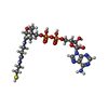  Mass: 687.554 Da / Num. of mol.: 1 / Source method: obtained synthetically / Formula: C21H35N7O13P2S / Feature type: SUBJECT OF INVESTIGATION Mass: 687.554 Da / Num. of mol.: 1 / Source method: obtained synthetically / Formula: C21H35N7O13P2S / Feature type: SUBJECT OF INVESTIGATION |
| #4: Water | ChemComp-HOH /  Mass: 18.015 Da / Num. of mol.: 22 / Source method: isolated from a natural source / Formula: H2O Mass: 18.015 Da / Num. of mol.: 22 / Source method: isolated from a natural source / Formula: H2O |
| Has ligand of interest | Y |
-Experimental details
-Experiment
| Experiment | Method: X-RAY DIFFRACTION / Number of used crystals: 1 |
|---|
- Sample preparation
| Crystal | Density Matthews: 5.64 Å3/Da / Density % sol: 78.21 % |
|---|---|
| Crystal grow | Temperature: 298 K / Method: vapor diffusion, hanging drop / pH: 5.6 Details: 1M LITHIUM SULPHATE, 2M AMMONIUM SULPHATE, 0.1M SODIUM CITRATE, PH 5.6 |
-Data collection
| Diffraction | Mean temperature: 100 K / Serial crystal experiment: N |
|---|---|
| Diffraction source | Source: SYNCHROTRON / Site: RRCAT INDUS-2  / Beamline: PX-BL21 / Wavelength: 1 Å / Beamline: PX-BL21 / Wavelength: 1 Å |
| Detector | Type: MARMOSAIC 225 mm CCD / Detector: CCD / Date: Jul 10, 2019 |
| Radiation | Protocol: SINGLE WAVELENGTH / Monochromatic (M) / Laue (L): M / Scattering type: x-ray |
| Radiation wavelength | Wavelength: 1 Å / Relative weight: 1 |
| Reflection | Resolution: 3.2→49.5 Å / Num. obs: 6957 / % possible obs: 92.27 % / Redundancy: 8.5 % / CC1/2: 0.98 / Rrim(I) all: 0.117 / Net I/σ(I): 30.61 |
| Reflection shell | Resolution: 3.2→3.28 Å / Redundancy: 13 % / Mean I/σ(I) obs: 5.02 / Num. unique obs: 534 / CC1/2: 0.92 / Rrim(I) all: 0.7 |
- Processing
| Software |
| ||||||||||||||||||||||||||||||||||||||||||||||||||||||||||||||||||||||||||||||||||||||||||||||||||||||||||||||||||||||||||||||||||||||||||||||||||||||
|---|---|---|---|---|---|---|---|---|---|---|---|---|---|---|---|---|---|---|---|---|---|---|---|---|---|---|---|---|---|---|---|---|---|---|---|---|---|---|---|---|---|---|---|---|---|---|---|---|---|---|---|---|---|---|---|---|---|---|---|---|---|---|---|---|---|---|---|---|---|---|---|---|---|---|---|---|---|---|---|---|---|---|---|---|---|---|---|---|---|---|---|---|---|---|---|---|---|---|---|---|---|---|---|---|---|---|---|---|---|---|---|---|---|---|---|---|---|---|---|---|---|---|---|---|---|---|---|---|---|---|---|---|---|---|---|---|---|---|---|---|---|---|---|---|---|---|---|---|---|---|---|
| Refinement | Method to determine structure: MOLECULAR REPLACEMENT Starting model: 6JOG Resolution: 3.202→49.497 Å / Cor.coef. Fo:Fc: 0.952 / Cor.coef. Fo:Fc free: 0.927 / SU B: 13.406 / SU ML: 0.221 / Cross valid method: THROUGHOUT / ESU R: 0.643 / ESU R Free: 0.354 Details: Hydrogens have been added in their riding positions
| ||||||||||||||||||||||||||||||||||||||||||||||||||||||||||||||||||||||||||||||||||||||||||||||||||||||||||||||||||||||||||||||||||||||||||||||||||||||
| Solvent computation | Ion probe radii: 0.8 Å / Shrinkage radii: 0.8 Å / VDW probe radii: 1.2 Å | ||||||||||||||||||||||||||||||||||||||||||||||||||||||||||||||||||||||||||||||||||||||||||||||||||||||||||||||||||||||||||||||||||||||||||||||||||||||
| Displacement parameters | Biso mean: 76.279 Å2
| ||||||||||||||||||||||||||||||||||||||||||||||||||||||||||||||||||||||||||||||||||||||||||||||||||||||||||||||||||||||||||||||||||||||||||||||||||||||
| Refinement step | Cycle: LAST / Resolution: 3.202→49.497 Å
| ||||||||||||||||||||||||||||||||||||||||||||||||||||||||||||||||||||||||||||||||||||||||||||||||||||||||||||||||||||||||||||||||||||||||||||||||||||||
| Refine LS restraints |
| ||||||||||||||||||||||||||||||||||||||||||||||||||||||||||||||||||||||||||||||||||||||||||||||||||||||||||||||||||||||||||||||||||||||||||||||||||||||
| LS refinement shell |
|
