 Movie
Movie Controller

[English] 日本語
 Yorodumi
- PDB-6kkh: Crystal structure of the oxalate bound malyl-CoA lyase from Rosei... -
+ Open data

- Basic information
| Entry | Database: PDB / ID: 6kkh | |||||||||
|---|---|---|---|---|---|---|---|---|---|---|
| Title | Crystal structure of the oxalate bound malyl-CoA lyase from Roseiflexus castenholzii | |||||||||
 Components Components | HpcH/HpaI aldolase | |||||||||
 Keywords Keywords | LYASE / Malyl-CoA lyase / CitE-like superfamily / Roseiflexus castenholzii / Conformational changes | |||||||||
| Function / homology |  Function and homology information Function and homology informationoxaloacetate metabolic process / catalytic activity / magnesium ion binding Similarity search - Function | |||||||||
| Biological species |  Roseiflexus castenholzii (bacteria) Roseiflexus castenholzii (bacteria) | |||||||||
| Method |  X-RAY DIFFRACTION / SYNCHROTRON / MOLECULAR REPLACEMENT / Resolution: 2.64 Å X-RAY DIFFRACTION / SYNCHROTRON / MOLECULAR REPLACEMENT / Resolution: 2.64 Å | |||||||||
 Authors Authors | Tang, W.R. / Wang, Z.G. / Zhang, C.Y. / Wang, C. | |||||||||
| Funding support |  China, 2items China, 2items
| |||||||||
 Citation Citation | Journal: Biochem.Biophys.Res.Commun. / Year: 2019 Title: The C-terminal domain conformational switch revealed by the crystal structure of malyl-CoA lyase from Roseiflexus castenholzii. Authors: Tang, W. / Wang, Z. / Zhang, C. / Wang, C. / Min, Z. / Zhang, X. / Liu, D. / Shen, J. / Xu, X. | |||||||||
| History |
|
- Structure visualization
| Structure viewer | Molecule: MolmilJmol/JSmol |
|---|
- Downloads & links
-Download
| PDBx/mmCIF format | 6kkh.cif.gz | 757.8 KB | Display | PDBx/mmCIF format |
|---|---|---|---|---|
| PDB format | pdb6kkh.ent.gz | 631.2 KB | Display | PDB format |
| PDBx/mmJSON format | 6kkh.json.gz | Tree view | PDBx/mmJSON format | |
| Others |  Other downloads Other downloads |
-Validation report
| Arichive directory | https://data.pdbj.org/pub/pdb/validation_reports/kk/6kkhftp://data.pdbj.org/pub/pdb/validation_reports/kk/6kkh | HTTPS FTP |
|---|
-Related structure data
| Related structure data |  6kinSC S: Starting model for refinement C: citing same article ( |
|---|---|
| Similar structure data |


-Links
 PDBj
PDBj
- Assembly
| Deposited unit | 
| ||||||||
|---|---|---|---|---|---|---|---|---|---|
| 1 | 
| ||||||||
| 2 | 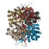
| ||||||||
| Unit cell |
|
-Components
| #1: Protein | Mass: 38316.215 Da / Num. of mol.: 12 Source method: isolated from a genetically manipulated source Source: (gene. exp.) Roseiflexus castenholzii (strain DSM 13941 / HLO8) (bacteria)Strain: DSM 13941 / HLO8 / Gene: Rcas_0912 / Production host: #2: Chemical | ChemComp-TRS /   Mass: 122.143 Da / Num. of mol.: 4 / Source method: obtained synthetically / Formula: C4H12NO3 / Comment: pH buffer*YM Mass: 122.143 Da / Num. of mol.: 4 / Source method: obtained synthetically / Formula: C4H12NO3 / Comment: pH buffer*YM#3: Chemical | ChemComp-OXL /   Mass: 88.019 Da / Num. of mol.: 4 / Source method: obtained synthetically / Formula: C2O4 / Feature type: SUBJECT OF INVESTIGATION Mass: 88.019 Da / Num. of mol.: 4 / Source method: obtained synthetically / Formula: C2O4 / Feature type: SUBJECT OF INVESTIGATION#4: Chemical |   Mass: 24.305 Da / Num. of mol.: 2 / Source method: obtained synthetically / Formula: Mg / Feature type: SUBJECT OF INVESTIGATION Mass: 24.305 Da / Num. of mol.: 2 / Source method: obtained synthetically / Formula: Mg / Feature type: SUBJECT OF INVESTIGATION#5: Water | ChemComp-HOH / |  Mass: 18.015 Da / Num. of mol.: 272 / Source method: isolated from a natural source / Formula: H2O Mass: 18.015 Da / Num. of mol.: 272 / Source method: isolated from a natural source / Formula: H2OHas ligand of interest | Y | |
|---|
-Experimental details
-Experiment
| Experiment | Method: X-RAY DIFFRACTION / Number of used crystals: 1 |
|---|
- Sample preparation
| Crystal | Density Matthews: 2.61 Å3/Da / Density % sol: 52.79 % |
|---|---|
| Crystal grow | Temperature: 289 K / Method: vapor diffusion, hanging drop / pH: 8 Details: The RfxMCL was incubated with Maganesium ion, oxalate and propionyl-CoA at a 1:1.5:1.5 molar ratio before crystallization to obtain the crystal of RfxMCL-OXL in a reservoir solution ...Details: The RfxMCL was incubated with Maganesium ion, oxalate and propionyl-CoA at a 1:1.5:1.5 molar ratio before crystallization to obtain the crystal of RfxMCL-OXL in a reservoir solution containing 16 % PEG8000, 0.2 M ammonium acetate and 0.1 M Tris pH8.0. |
-Data collection
| Diffraction | Mean temperature: 100 K / Serial crystal experiment: N |
|---|---|
| Diffraction source | Source: SYNCHROTRON / Site: SSRF / Beamline: BL19U1 / Wavelength: 0.97891 Å |
| Detector | Type: ADSC QUANTUM 315r / Detector: CCD / Date: Dec 11, 2018 |
| Radiation | Protocol: SINGLE WAVELENGTH / Monochromatic (M) / Laue (L): M / Scattering type: x-ray |
| Radiation wavelength | Wavelength: 0.97891 Å / Relative weight: 1 |
| Reflection | Resolution: 2.64→50 Å / Num. obs: 133993 / % possible obs: 99 % / Redundancy: 6.3 % / CC1/2: 0.995 / Rmerge(I) obs: 0.063 / Rpim(I) all: 0.038 / Rrim(I) all: 0.097 / Net I/σ(I): 16.3 |
| Reflection shell | Resolution: 2.64→2.7 Å / Redundancy: 6.1 % / Mean I/σ(I) obs: 2.21 / Num. unique obs: 6605 / CC1/2: 0.879 / Rsym value: 0.689 / % possible all: 98.5 |
- Processing
| Software |
| ||||||||||||||||||||||||||||||||||||||||||||||||||||||||||||
|---|---|---|---|---|---|---|---|---|---|---|---|---|---|---|---|---|---|---|---|---|---|---|---|---|---|---|---|---|---|---|---|---|---|---|---|---|---|---|---|---|---|---|---|---|---|---|---|---|---|---|---|---|---|---|---|---|---|---|---|---|---|
| Refinement | Method to determine structure: MOLECULAR REPLACEMENT Starting model: 6KIN Resolution: 2.64→49.48 Å / Cor.coef. Fo:Fc: 0.943 / Cor.coef. Fo:Fc free: 0.904 / SU B: 14.101 / SU ML: 0.291 / Cross valid method: FREE R-VALUE / σ(F): 0 / ESU R Free: 0.35 Details: HYDROGENS HAVE BEEN ADDED IN THE RIDING POSITIONS U VALUES : REFINED INDIVIDUALLY
| ||||||||||||||||||||||||||||||||||||||||||||||||||||||||||||
| Solvent computation | Ion probe radii: 0.8 Å / Shrinkage radii: 0.8 Å / VDW probe radii: 1.2 Å | ||||||||||||||||||||||||||||||||||||||||||||||||||||||||||||
| Displacement parameters | Biso max: 136.17 Å2 / Biso mean: 56.437 Å2 / Biso min: 14.85 Å2
| ||||||||||||||||||||||||||||||||||||||||||||||||||||||||||||
| Refinement step | Cycle: final / Resolution: 2.64→49.48 Å
| ||||||||||||||||||||||||||||||||||||||||||||||||||||||||||||
| Refine LS restraints |
| ||||||||||||||||||||||||||||||||||||||||||||||||||||||||||||
| LS refinement shell | Resolution: 2.64→2.709 Å / Rfactor Rfree error: 0 / Total num. of bins used: 20
|
