 Movie
Movie Controller


+ Open data

- Basic information
| Entry | Database: PDB / ID: 6kjg | ||||||
|---|---|---|---|---|---|---|---|
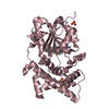
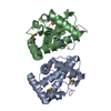
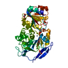
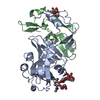
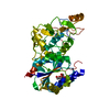
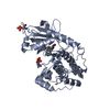
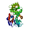
| Title | Crystal structure of PsoF | ||||||
 Components Components | Dual-functional monooxygenase/methyltransferase psoF | ||||||
 Keywords Keywords | OXIDOREDUCTASE / metyltransferase | ||||||
| Function / homology |  Function and homology information Function and homology informationfumagillin biosynthetic process / N,N-dimethylaniline monooxygenase activity / Oxidoreductases / secondary metabolite biosynthetic process / Transferases; Transferring one-carbon groups; Methyltransferases / methyltransferase activity / monooxygenase activity / NADP binding / flavin adenine dinucleotide binding / methylation Similarity search - Function | ||||||
| Biological species |  | ||||||
| Method |  X-RAY DIFFRACTION / SYNCHROTRON / MOLECULAR REPLACEMENT / Resolution: 1.99 Å X-RAY DIFFRACTION / SYNCHROTRON / MOLECULAR REPLACEMENT / Resolution: 1.99 Å | ||||||
 Authors Authors | Hara, K. / Hashimoto, H. / Matsushita, T. / Tsunematsu, Y. / Watanabe, K. | ||||||
 Citation Citation | Journal: Biochemistry / Year: 2019 Title: Functional and Structural Analyses oftrans C-Methyltransferase in Fungal Polyketide Biosynthesis. Authors: Kishimoto, S. / Tsunematsu, Y. / Matsushita, T. / Hara, K. / Hashimoto, H. / Tang, Y. / Watanabe, K. | ||||||
| History |
|
- Structure visualization
| Structure viewer | Molecule: MolmilJmol/JSmol |
|---|
- Downloads & links
-Download
| PDBx/mmCIF format | 6kjg.cif.gz | 221.2 KB | Display | PDBx/mmCIF format |
|---|---|---|---|---|
| PDB format | pdb6kjg.ent.gz | 175.6 KB | Display | PDB format |
| PDBx/mmJSON format | 6kjg.json.gz | Tree view | PDBx/mmJSON format | |
| Others |  Other downloads Other downloads |
-Validation report
| Arichive directory | https://data.pdbj.org/pub/pdb/validation_reports/kj/6kjgftp://data.pdbj.org/pub/pdb/validation_reports/kj/6kjg | HTTPS FTP |
|---|
-Related structure data
| Related structure data |  6kjiC 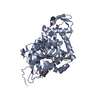 5thyS S: Starting model for refinement C: citing same article ( |
|---|---|
| Similar structure data |


-Links
 PDBj
PDBj- Assembly
| Deposited unit | 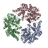
| ||||||||||||
|---|---|---|---|---|---|---|---|---|---|---|---|---|---|
| 1 | 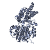
| ||||||||||||
| 2 | 
| ||||||||||||
| 3 | 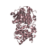
| ||||||||||||
| Unit cell |
| ||||||||||||
| Components on special symmetry positions |
|
-Components
| #1: Protein | Mass: 41732.039 Da / Num. of mol.: 3 Source method: isolated from a genetically manipulated source Source: (gene. exp.) Strain: ATCC MYA-4609 / Af293 / CBS 101355 / FGSC A1100 / Gene: psoF, AFUA_8G00440 / Production host:  References: UniProt: Q4WAZ0, Oxidoreductases, Transferases; Transferring one-carbon groups; Methyltransferases #2: Water | ChemComp-HOH / |  Mass: 18.015 Da / Num. of mol.: 487 / Source method: isolated from a natural source / Formula: H2O Mass: 18.015 Da / Num. of mol.: 487 / Source method: isolated from a natural source / Formula: H2O |
|---|
-Experimental details
-Experiment
| Experiment | Method: X-RAY DIFFRACTION / Number of used crystals: 1 |
|---|
- Sample preparation
| Crystal | Density Matthews: 2.57 Å3/Da / Density % sol: 52.1 % |
|---|---|
| Crystal grow | Temperature: 293 K / Method: vapor diffusion, hanging drop / Details: PEG 3350, sodium fluoride, bis-tris propane pH 6.5 |
-Data collection
| Diffraction | Mean temperature: 100 K / Serial crystal experiment: N |
|---|---|
| Diffraction source | Source: SYNCHROTRON / Site: Photon Factory  / Beamline: BL-17A / Wavelength: 0.98 Å / Beamline: BL-17A / Wavelength: 0.98 Å |
| Detector | Type: DECTRIS PILATUS3 S 6M / Detector: PIXEL / Date: Nov 11, 2017 |
| Radiation | Protocol: SINGLE WAVELENGTH / Monochromatic (M) / Laue (L): M / Scattering type: x-ray |
| Radiation wavelength | Wavelength: 0.98 Å / Relative weight: 1 |
| Reflection | Resolution: 1.99→20 Å / Num. obs: 80787 / % possible obs: 98.3 % / Redundancy: 3 % / Rmerge(I) obs: 0.054 / Net I/σ(I): 11.87 |
| Reflection shell | Resolution: 1.99→2.09 Å / Rmerge(I) obs: 0.332 / Mean I/σ(I) obs: 2.48 / Num. unique obs: 12540 / % possible all: 95.8 |
- Processing
| Software |
| ||||||||||||||||||||
|---|---|---|---|---|---|---|---|---|---|---|---|---|---|---|---|---|---|---|---|---|---|
| Refinement | Method to determine structure: MOLECULAR REPLACEMENT Starting model: 5THY Resolution: 1.99→19.8 Å / Cross valid method: FREE R-VALUE
| ||||||||||||||||||||
| Refinement step | Cycle: LAST / Resolution: 1.99→19.8 Å
|
