 Movie
Movie Controller


+ Open data

- Basic information
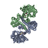
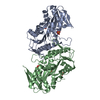
| Entry | Database: PDB / ID: 1b5f | ||||||||||||
|---|---|---|---|---|---|---|---|---|---|---|---|---|---|
| Title | NATIVE CARDOSIN A FROM CYNARA CARDUNCULUS L. | ||||||||||||
 Components Components | (PROTEIN (CARDOSIN ...) x 2 | ||||||||||||
 Keywords Keywords | HYDROLASE / ASPARTIC PROTEINASE | ||||||||||||
| Function / homology |  Function and homology information Function and homology informationprotein storage vacuole / Hydrolases; Acting on peptide bonds (peptidases); Aspartic endopeptidases / lipid metabolic process / aspartic-type endopeptidase activity / proteolysis Similarity search - Function | ||||||||||||
| Biological species |  Cynara cardunculus (cardoon) Cynara cardunculus (cardoon) | ||||||||||||
| Method |  X-RAY DIFFRACTION / SYNCHROTRON / MOLECULAR REPLACEMENT / Resolution: 1.72 Å X-RAY DIFFRACTION / SYNCHROTRON / MOLECULAR REPLACEMENT / Resolution: 1.72 Å | ||||||||||||
 Authors Authors | Frazao, C. / Bento, I. / Carrondo, M.A. | ||||||||||||
 Citation Citation | Journal: J.Biol.Chem. / Year: 1999 Title: Crystal structure of cardosin A, a glycosylated and Arg-Gly-Asp-containing aspartic proteinase from the flowers of Cynara cardunculus L. Authors: Frazao, C. / Bento, I. / Costa, J. / Soares, C.M. / Verissimo, P. / Faro, C. / Pires, E. / Cooper, J. / Carrondo, M.A. #1: Journal: Acta Crystallogr.,Sect.D / Year: 1999Title: Crystallization and Preliminary X-Ray Crystallographic Studies of the Plant Aspartic Proteinase Cardosin A Authors: Bento, I. / Frazao, C. / Coelho, R. / Wilson, K. / Dauter, Z. / Carrondo, M.A. #2: Journal: Adv.Exp.Med.Biol. / Year: 1998Title: Crystallization, Structure Solution, and Initial Refinement of Plant Cardosin-A Authors: Bento, I. / Coelho, R. / Frazao, C. / Costa, J. / Faro, C. / Verissimo, P. / Pires, E. / Cooper, J. / Dauter, Z. / Wilson, K. / Carrondo, M.A. | ||||||||||||
| History |
|
- Structure visualization
| Structure viewer | Molecule: MolmilJmol/JSmol |
|---|
- Downloads & links
-Download
| PDBx/mmCIF format | 1b5f.cif.gz | 159.4 KB | Display | PDBx/mmCIF format |
|---|---|---|---|---|
| PDB format | pdb1b5f.ent.gz | 124.3 KB | Display | PDB format |
| PDBx/mmJSON format | 1b5f.json.gz | Tree view | PDBx/mmJSON format | |
| Others |  Other downloads Other downloads |
-Validation report
| Arichive directory | https://data.pdbj.org/pub/pdb/validation_reports/b5/1b5fftp://data.pdbj.org/pub/pdb/validation_reports/b5/1b5f | HTTPS FTP |
|---|
-Related structure data
| Related structure data | 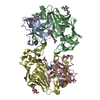 1lybS S: Starting model for refinement |
|---|---|
| Similar structure data |


-Links
 PDBj
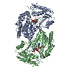
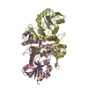
PDBj- Assembly
| Deposited unit | 
| ||||||||
|---|---|---|---|---|---|---|---|---|---|
| 1 |
| ||||||||
| 2 | 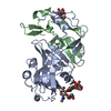
| ||||||||
| 3 | 
| ||||||||
| Unit cell |
| ||||||||
| Components on special symmetry positions |
| ||||||||
| Noncrystallographic symmetry (NCS) | NCS oper: (Code: given Matrix: (-0.88365, -0.13686, 0.44769), Vector: |
-Components
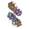
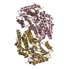
-PROTEIN (CARDOSIN ... , 2 types, 4 molecules ACBD
| #1: Protein | Mass: 26042.812 Da / Num. of mol.: 2 / Source method: isolated from a natural source Details: RESIDUE NAMES ACCORDING TO PEPSIN NUMBERING AFTER A.R.SIELECKI, A.A.FEDOROV, A.BOODHOO, N.S.ANDREEVA, AND M.N.G.JAMES (1990).J.MOL.BIOL. 214, 143-170. NATIVE CARDOSIN A SEQUENCE DIFFERS FROM ...Details: RESIDUE NAMES ACCORDING TO PEPSIN NUMBERING AFTER A.R.SIELECKI, A.A.FEDOROV, A.BOODHOO, N.S.ANDREEVA, AND M.N.G.JAMES (1990).J.MOL.BIOL. 214, 143-170. NATIVE CARDOSIN A SEQUENCE DIFFERS FROM THAT DEDUCED FROM CDNA THROUGH EXCISION OF THE PSI DOMAIN. MATURE CARDOSIN A IS FOUND IN A TWO CHAIN FORM DUE TO A POST-TRANSLATIONAL CLEAVAGE EVENT. A FIRST, 35 KD CHAIN COMPRISES RESIDUES 0/1 - 238 AND THE SECOND 15 KD CHAIN COMPRISES RESIDUES 243 - 326. THE ASYMMETRIC UNIT CONTAINS TWO CARDOSIN A MOLECULES. MOLECULE 1 HAS BEEN ASSIGNED CHAIN INDICATORS *A* AND *B*, AND MOLECULE 2 HAVE BEEN ASSIGNED CHAIN INDICATORS *C* AND *D*. MOLECULE 1 COMPOSED BY CHAINS A(0-238) AND B (243-326). MOLECULE 2 COMPOSED BY CHAINS C(1-238) AND D (243-326). N-LINKED CARBOHYDRATES (RESIDUES 401-405 A AND 401-406 C) ATTACHED TO ASN 67. N-LINKED CARBOHYDRATES (RESIDUES 501-504 B AND 501-504 D) ATTACHED TO ASN 257. Source: (natural) Cynara cardunculus (cardoon) / Organ: FLOWER;PISTIL / Organelle: STORAGE VACUOLES / Tissue: PAPILLAR EPIDERMIS OF THE STIGMAReferences: EMBL: CAB4134, UniProt: Q9XFX3*PLUS, Hydrolases; Acting on peptide bonds (peptidases); Aspartic endopeptidases #2: Protein | Mass: 9441.812 Da / Num. of mol.: 2 / Source method: isolated from a natural source Details: RESIDUE NAMES ACCORDING TO PEPSIN NUMBERING AFTER A.R.SIELECKI, A.A.FEDOROV, A.BOODHOO, N.S.ANDREEVA, AND M.N.G.JAMES (1990).J.MOL.BIOL. 214, 143-170. NATIVE CARDOSIN A SEQUENCE DIFFERS FROM ...Details: RESIDUE NAMES ACCORDING TO PEPSIN NUMBERING AFTER A.R.SIELECKI, A.A.FEDOROV, A.BOODHOO, N.S.ANDREEVA, AND M.N.G.JAMES (1990).J.MOL.BIOL. 214, 143-170. NATIVE CARDOSIN A SEQUENCE DIFFERS FROM THAT DEDUCED FROM CDNA THROUGH EXCISION OF THE PSI DOMAIN. MATURE CARDOSIN A IS FOUND IN A TWO CHAIN FORM DUE TO A POST-TRANSLATIONAL CLEAVAGE EVENT. A FIRST, 35 KD CHAIN COMPRISES RESIDUES 0/1 - 238 AND THE SECOND 15 KD CHAIN COMPRISES RESIDUES 243 - 326. THE ASYMMETRIC UNIT CONTAINS TWO CARDOSIN A MOLECULES. MOLECULE 1 HAS BEEN ASSIGNED CHAIN INDICATORS *A* AND *B*, AND MOLECULE 2 HAVE BEEN ASSIGNED CHAIN INDICATORS *C* AND *D*. MOLECULE 1 COMPOSED BY CHAINS A(0-238) AND B (243-326). MOLECULE 2 COMPOSED BY CHAINS C(1-238) AND D (243-326). N-LINKED CARBOHYDRATES (RESIDUES 401-405 A AND 401-406 C) ATTACHED TO ASN 67. N-LINKED CARBOHYDRATES (RESIDUES 501-504 B AND 501-504 D) ATTACHED TO ASN 257. Source: (natural) Cynara cardunculus (cardoon) / Organ: FLOWER;PISTIL / Organelle: STORAGE VACUOLES / Tissue: PAPILLAR EPIDERMIS OF THE STIGMAReferences: EMBL: CAB4134, UniProt: Q9XFX3*PLUS, Hydrolases; Acting on peptide bonds (peptidases); Aspartic endopeptidases |
|---|
-Sugars , 3 types, 4 molecules
| #3: Polysaccharide | alpha-D-mannopyranose-(1-3)-beta-D-mannopyranose-(1-4)-2-acetamido-2-deoxy-beta-D-glucopyranose-(1- ...alpha-D-mannopyranose-(1-3)-beta-D-mannopyranose-(1-4)-2-acetamido-2-deoxy-beta-D-glucopyranose-(1-4)-[alpha-L-fucopyranose-(1-3)]2-acetamido-2-deoxy-beta-D-glucopyranose Source method: isolated from a genetically manipulated source | ||
|---|---|---|---|
| #4: Polysaccharide | Source method: isolated from a genetically manipulated source #5: Polysaccharide | alpha-D-mannopyranose-(1-3)-[alpha-D-mannopyranose-(1-6)]beta-D-mannopyranose-(1-4)-2-acetamido-2- ...alpha-D-mannopyranose-(1-3)-[alpha-D-mannopyranose-(1-6)]beta-D-mannopyranose-(1-4)-2-acetamido-2-deoxy-beta-D-glucopyranose-(1-4)-[alpha-L-fucopyranose-(1-3)]2-acetamido-2-deoxy-beta-D-glucopyranose | Source method: isolated from a genetically manipulated source |
-Non-polymers , 1 types, 528 molecules 
| #6: Water | ChemComp-HOH / Mass: 18.015 Da / Num. of mol.: 528 / Source method: isolated from a natural source / Formula: H2O |
|---|
-Details
| Compound details | MATURE CARDOSIN A IS FOUND IN A TWO CHAIN FORM DUE TO A POST-TRANSLATIONAL CLEAVAGE EVENT. A FIRST, ...MATURE CARDOSIN A IS FOUND IN A TWO CHAIN FORM DUE TO A POST-TRANSLATIO |
|---|---|
| Has protein modification | Y |
-Experimental details
-Experiment
| Experiment | Method: X-RAY DIFFRACTION / Number of used crystals: 1 |
|---|
- Sample preparation
| Crystal | Density Matthews: 2.26 Å3/Da / Density % sol: 46 % | ||||||||||||||||||||||||||||||
|---|---|---|---|---|---|---|---|---|---|---|---|---|---|---|---|---|---|---|---|---|---|---|---|---|---|---|---|---|---|---|---|
| Crystal grow | pH: 5.5 Details: LYOPHILIZED PROTEIN DISSOLVED IN WATER TO 12 MG/ML, SITTING DROPS OF PROTEIN SOLUTION AN ALIQUOTA OF PRECIPITANT SOLUTION, COMPOSED OF PEG 4 K 40%, SODIUM CITRATE BUFFER 0.1 M AND AMMONIUM ACETATE 0.2 M, pH 5.5 | ||||||||||||||||||||||||||||||
| Crystal | *PLUS | ||||||||||||||||||||||||||||||
| Crystal grow | *PLUS Method: vapor diffusion, sitting drop / Details: Bento, I., (1998) Acta Cryst., D54, 991. | ||||||||||||||||||||||||||||||
| Components of the solutions | *PLUS
|
-Data collection
| Diffraction | Mean temperature: 100 K |
|---|---|
| Diffraction source | Source: SYNCHROTRON / Site: EMBL/DESY, HAMBURG  / Beamline: X11 / Wavelength: 0.9091 / Beamline: X11 / Wavelength: 0.9091 |
| Detector | Type: MARRESEARCH / Detector: IMAGE PLATE / Date: Apr 5, 1997 |
| Radiation | Protocol: SINGLE WAVELENGTH / Monochromatic (M) / Laue (L): M / Scattering type: x-ray |
| Radiation wavelength | Wavelength: 0.9091 Å / Relative weight: 1 |
| Reflection | Resolution: 1.72→34.78 Å / Num. obs: 84399 / % possible obs: 96.9 % / Redundancy: 11.3 % / Rmerge(I) obs: 0.062 / Net I/σ(I): 15.9 |
| Reflection shell | Resolution: 1.72→1.75 Å / Redundancy: 2.9 % / Rmerge(I) obs: 0.413 / Mean I/σ(I) obs: 2.5 / % possible all: 99.3 |
| Reflection shell | *PLUS % possible obs: 99.3 % |
- Processing
| Software |
| |||||||||||||||||||||||||||||||||
|---|---|---|---|---|---|---|---|---|---|---|---|---|---|---|---|---|---|---|---|---|---|---|---|---|---|---|---|---|---|---|---|---|---|---|
| Refinement | Method to determine structure: MOLECULAR REPLACEMENT Starting model: HUMAN CATHEPSIN D (PDB ENTRY 1LYB)) Resolution: 1.72→34.8 Å / Num. parameters: 23395 / Num. restraintsaints: 27500 / Cross valid method: FREE R / σ(F): 0 StereochEM target val spec case: FUCOSE STEREOCHEMICAL TARGET PARAMETERS DERIVED FROM 1.6 A RESOLUTION STRUCTURE PDB ENTRY 2MYR; MANNOSE STEREOCHEMICAL TARGET PARAMETERS DERIVED FROM 1.25 A ...StereochEM target val spec case: FUCOSE STEREOCHEMICAL TARGET PARAMETERS DERIVED FROM 1.6 A RESOLUTION STRUCTURE PDB ENTRY 2MYR; MANNOSE STEREOCHEMICAL TARGET PARAMETERS DERIVED FROM 1.25 A RESOLUTION STRUCTURE PDB ENTRY 2WEA; N- ACETYL-GLUCOSAMINE STEREOCHEMICAL TARGET PARAMETERS DERIVED FROM 1.5 A STRUCTURE PDB ENTRY 1LZB Stereochemistry target values: ENGH AND HUBER Details: ANISOTROPIC SCALING APPLIED BY THE METHOD OF PARKIN, MOEZZI & HOPE, J.APPL.CRYST.28 (1995)53-56. DISORDERED REGIONS THAT WERE MODELED STEREOCHEMICALLY: 75-78 CHAIN A AND 75-79 CHAIN C (FLAP); ...Details: ANISOTROPIC SCALING APPLIED BY THE METHOD OF PARKIN, MOEZZI & HOPE, J.APPL.CRYST.28 (1995)53-56. DISORDERED REGIONS THAT WERE MODELED STEREOCHEMICALLY: 75-78 CHAIN A AND 75-79 CHAIN C (FLAP); 46-46 CHAIN A AND 46-47 CHAIN C; 159-160 AND 160B CHAIN A. DISORDERED REGIONS THAT WERE MODELED STEREOCHEMICALLY: 75-78 CHAIN A AND 75-79 CHAIN C (FLAP); 46-46 CHAIN A AND 46-47 CHAIN C; 159-160 AND 160B CHAIN A.
| |||||||||||||||||||||||||||||||||
| Solvent computation | Solvent model: MOEWS & KRETSINGER, J.MOL.BIOL.91(1973)201-2 | |||||||||||||||||||||||||||||||||
| Refine analyze | Num. disordered residues: 18 / Occupancy sum hydrogen: 4964 / Occupancy sum non hydrogen: 5754.5 | |||||||||||||||||||||||||||||||||
| Refinement step | Cycle: LAST / Resolution: 1.72→34.8 Å
| |||||||||||||||||||||||||||||||||
| Refine LS restraints |
| |||||||||||||||||||||||||||||||||
| Software | *PLUS Name: SHELXL-97 / Classification: refinement | |||||||||||||||||||||||||||||||||
| Refinement | *PLUS σ(F): 0 / % reflection Rfree: 4.8 % / Rfactor Rwork: 0.205 | |||||||||||||||||||||||||||||||||
| Solvent computation | *PLUS | |||||||||||||||||||||||||||||||||
| Displacement parameters | *PLUS | |||||||||||||||||||||||||||||||||
| Refine LS restraints | *PLUS Type: s_plane_restr / Dev ideal: 0.03 |
