 Movie
Movie Controller

[English] 日本語
 Yorodumi
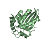
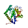
- PDB-6kis: High-resolution structure of mouse CXorf40A (soaked with Methylme... -
+ Open data

- Basic information
| Entry | Database: PDB / ID: 6kis | ||||||
|---|---|---|---|---|---|---|---|
| Title | High-resolution structure of mouse CXorf40A (soaked with Methylmercury chloride) | ||||||
 Components Components | Uncharacterized protein CXorf40 homolog | ||||||
 Keywords Keywords | IMMUNE SYSTEM / PUA-like / ASCH domain / Chromosome X / mitochondria | ||||||
| Function / homology | Protein EOLA1/EOLA2 / ASCH / ASCH domain / ASCH domain / PUA-like superfamily / : / Protein EOLA1 Function and homology information Function and homology information | ||||||
| Biological species |  | ||||||
| Method |  X-RAY DIFFRACTION / SYNCHROTRON / SAD / Resolution: 1.54 Å X-RAY DIFFRACTION / SYNCHROTRON / SAD / Resolution: 1.54 Å | ||||||
 Authors Authors | Wu, B.X. | ||||||
 Citation Citation | Journal: To Be Published Title: High-resolution structure of mouse CXorf40A (soaked with Methylmercury chloride) Authors: Wu, B.X. | ||||||
| History |
|
- Structure visualization
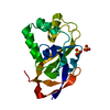
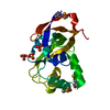
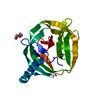
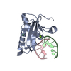
| Structure viewer | Molecule: MolmilJmol/JSmol |
|---|
- Downloads & links
-Download
| PDBx/mmCIF format | 6kis.cif.gz | 49.5 KB | Display | PDBx/mmCIF format |
|---|---|---|---|---|
| PDB format | pdb6kis.ent.gz | 33.8 KB | Display | PDB format |
| PDBx/mmJSON format | 6kis.json.gz | Tree view | PDBx/mmJSON format | |
| Others |  Other downloads Other downloads |
-Validation report
| Arichive directory | https://data.pdbj.org/pub/pdb/validation_reports/ki/6kisftp://data.pdbj.org/pub/pdb/validation_reports/ki/6kis | HTTPS FTP |
|---|
-Related structure data
| Related structure data | |
|---|---|
| Similar structure data |


-Links
 PDBj
PDBj


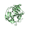
- Assembly
| Deposited unit | 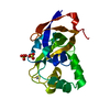
| ||||||||||||
|---|---|---|---|---|---|---|---|---|---|---|---|---|---|
| 1 |
| ||||||||||||
| Unit cell |
| ||||||||||||
| Components on special symmetry positions |
|
-Components
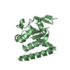
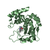
| #1: Protein | Mass: 18278.992 Da / Num. of mol.: 1 Source method: isolated from a genetically manipulated source Source: (gene. exp.)  | ||||||
|---|---|---|---|---|---|---|---|
| #2: Chemical |   Mass: 96.063 Da / Num. of mol.: 2 / Source method: obtained synthetically / Formula: SO4 Mass: 96.063 Da / Num. of mol.: 2 / Source method: obtained synthetically / Formula: SO4#3: Chemical | ChemComp-HG / |   Mass: 200.590 Da / Num. of mol.: 1 / Source method: obtained synthetically / Formula: Hg Mass: 200.590 Da / Num. of mol.: 1 / Source method: obtained synthetically / Formula: Hg#4: Water | ChemComp-HOH / |  Mass: 18.015 Da / Num. of mol.: 116 / Source method: isolated from a natural source / Formula: H2O Mass: 18.015 Da / Num. of mol.: 116 / Source method: isolated from a natural source / Formula: H2OHas ligand of interest | N | |
-Experimental details
-Experiment
| Experiment | Method: X-RAY DIFFRACTION / Number of used crystals: 1 |
|---|
- Sample preparation
| Crystal | Density Matthews: 2.23 Å3/Da / Density % sol: 44.8 % |
|---|---|
| Crystal grow | Temperature: 293 K / Method: vapor diffusion, sitting drop Details: 0.1 M HEPES sodium pH 7.5, 2% v/v Polyethylene glycol 400, 2.0 M Ammonium sulfate |
-Data collection
| Diffraction | Mean temperature: 100 K / Serial crystal experiment: N |
|---|---|
| Diffraction source | Source: SYNCHROTRON / Site: SSRF  / Beamline: BL17U1 / Wavelength: 1.00928 Å / Beamline: BL17U1 / Wavelength: 1.00928 Å |
| Detector | Type: ADSC QUANTUM 315r / Detector: CCD / Date: Jun 22, 2017 |
| Radiation | Protocol: SINGLE WAVELENGTH / Monochromatic (M) / Laue (L): M / Scattering type: x-ray |
| Radiation wavelength | Wavelength: 1.00928 Å / Relative weight: 1 |
| Reflection | Resolution: 1.54→50 Å / Num. obs: 24850 / % possible obs: 99.5 % / Redundancy: 10.2 % / Net I/σ(I): 36.57 |
| Reflection shell | Resolution: 1.54→1.6 Å / Num. unique obs: 2461 |
- Processing
| Software |
| ||||||||||||||||
|---|---|---|---|---|---|---|---|---|---|---|---|---|---|---|---|---|---|
| Refinement | Method to determine structure: SAD / Resolution: 1.54→30 Å / Cross valid method: FREE R-VALUE
| ||||||||||||||||
| Refinement step | Cycle: LAST / Resolution: 1.54→30 Å
|
