 Movie
Movie Controller

[English] 日本語
 Yorodumi
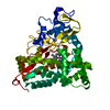
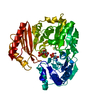
- PDB-6kfw: The cytochrome P450 enzyme CxnD for C-S bond formation in chuangx... -
+ Open data

- Basic information
| Entry | Database: PDB / ID: 6kfw | ||||||
|---|---|---|---|---|---|---|---|
| Title | The cytochrome P450 enzyme CxnD for C-S bond formation in chuangxinmycin biosynthesis | ||||||
 Components Components | CxnD | ||||||
 Keywords Keywords | OXIDOREDUCTASE / P450 / CxnD | ||||||
| Function / homology | Chem-D8L / PROTOPORPHYRIN IX CONTAINING FE Function and homology information Function and homology information | ||||||
| Biological species |  Actinoplanes tsinanensis (bacteria) Actinoplanes tsinanensis (bacteria) | ||||||
| Method |  X-RAY DIFFRACTION / SYNCHROTRON / MOLECULAR REPLACEMENT / Resolution: 2 Å X-RAY DIFFRACTION / SYNCHROTRON / MOLECULAR REPLACEMENT / Resolution: 2 Å | ||||||
 Authors Authors | Hong, B. | ||||||
 Citation Citation | Journal: Angew.Chem.Int.Ed.Engl. / Year: 2021 Title: The Cytochrome P450 Catalyzing C-S Bond Formation in S-Heterocyclization of Chuangxinmycin Biosynthesis. Authors: Shi, Y. / Jiang, Z. / Hu, X. / Hu, X. / Gu, R. / Jiang, B. / Zuo, L. / Li, X. / Sun, H. / Zhang, C. / Wang, L. / Wu, L. / Hong, B. | ||||||
| History |
|
- Structure visualization
| Structure viewer | Molecule: MolmilJmol/JSmol |
|---|
- Downloads & links
-Download
| PDBx/mmCIF format | 6kfw.cif.gz | 111.2 KB | Display | PDBx/mmCIF format |
|---|---|---|---|---|
| PDB format | pdb6kfw.ent.gz | 75.8 KB | Display | PDB format |
| PDBx/mmJSON format | 6kfw.json.gz | Tree view | PDBx/mmJSON format | |
| Others |  Other downloads Other downloads |
-Validation report
| Arichive directory | https://data.pdbj.org/pub/pdb/validation_reports/kf/6kfwftp://data.pdbj.org/pub/pdb/validation_reports/kf/6kfw | HTTPS FTP |
|---|
-Related structure data
| Related structure data |  2whwS S: Starting model for refinement |
|---|---|
| Similar structure data |


-Links
 PDBj
PDBj- Assembly
| Deposited unit | 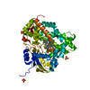
| ||||||||||||
|---|---|---|---|---|---|---|---|---|---|---|---|---|---|
| 1 |
| ||||||||||||
| Unit cell |
|
-Components
| #1: Protein | Mass: 45372.527 Da / Num. of mol.: 1 Source method: isolated from a genetically manipulated source Source: (gene. exp.) Actinoplanes tsinanensis (bacteria) / Production host: | ||||||
|---|---|---|---|---|---|---|---|
| #2: Chemical | ChemComp-HEM /   Mass: 616.487 Da / Num. of mol.: 1 / Source method: obtained synthetically / Formula: C34H32FeN4O4 Mass: 616.487 Da / Num. of mol.: 1 / Source method: obtained synthetically / Formula: C34H32FeN4O4 | ||||||
| #3: Chemical |   Mass: 96.063 Da / Num. of mol.: 3 / Source method: obtained synthetically / Formula: SO4 Mass: 96.063 Da / Num. of mol.: 3 / Source method: obtained synthetically / Formula: SO4#4: Chemical | ChemComp-D8L / ( | 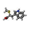  Mass: 235.302 Da / Num. of mol.: 1 / Source method: obtained synthetically / Formula: C12H13NO2S / Feature type: SUBJECT OF INVESTIGATION Mass: 235.302 Da / Num. of mol.: 1 / Source method: obtained synthetically / Formula: C12H13NO2S / Feature type: SUBJECT OF INVESTIGATION#5: Water | ChemComp-HOH / |  Mass: 18.015 Da / Num. of mol.: 278 / Source method: isolated from a natural source / Formula: H2O Mass: 18.015 Da / Num. of mol.: 278 / Source method: isolated from a natural source / Formula: H2OHas ligand of interest | Y | |
-Experimental details
-Experiment
| Experiment | Method: X-RAY DIFFRACTION / Number of used crystals: 1 |
|---|
- Sample preparation
| Crystal | Density Matthews: 2.42 Å3/Da / Density % sol: 49.21 % |
|---|---|
| Crystal grow | Temperature: 293 K / Method: vapor diffusion, hanging drop / Details: 1.8M ammonium sulfate |
-Data collection
| Diffraction | Mean temperature: 100 K / Serial crystal experiment: N |
|---|---|
| Diffraction source | Source: SYNCHROTRON / Site: SSRF  / Beamline: BL18U1 / Wavelength: 0.979 Å / Beamline: BL18U1 / Wavelength: 0.979 Å |
| Detector | Type: DECTRIS PILATUS 6M / Detector: PIXEL / Date: Mar 27, 2019 |
| Radiation | Protocol: SINGLE WAVELENGTH / Monochromatic (M) / Laue (L): M / Scattering type: x-ray |
| Radiation wavelength | Wavelength: 0.979 Å / Relative weight: 1 |
| Reflection | Resolution: 2→50 Å / Num. obs: 29784 / % possible obs: 100 % / Redundancy: 12.7 % / Biso Wilson estimate: 17.1 Å2 / Rmerge(I) obs: 0.175 / Rpim(I) all: 0.051 / Net I/σ(I): 13.9 |
| Reflection shell | Resolution: 2→2.03 Å / Redundancy: 10.8 % / Rmerge(I) obs: 0.634 / Mean I/σ(I) obs: 2.8 / Num. unique obs: 1312 / Rpim(I) all: 0.198 / % possible all: 100 |
- Processing
| Software |
| ||||||||||||||||||||||||||||||||||||||||||||||||||||||||||||||||||||||||||||||||||||
|---|---|---|---|---|---|---|---|---|---|---|---|---|---|---|---|---|---|---|---|---|---|---|---|---|---|---|---|---|---|---|---|---|---|---|---|---|---|---|---|---|---|---|---|---|---|---|---|---|---|---|---|---|---|---|---|---|---|---|---|---|---|---|---|---|---|---|---|---|---|---|---|---|---|---|---|---|---|---|---|---|---|---|---|---|---|
| Refinement | Method to determine structure: MOLECULAR REPLACEMENT Starting model: 2WHW Resolution: 2→25.88 Å / SU ML: 0.1981 / Cross valid method: FREE R-VALUE / σ(F): 1.38 / Phase error: 21.4137 / Stereochemistry target values: GeoStd + Monomer Library
| ||||||||||||||||||||||||||||||||||||||||||||||||||||||||||||||||||||||||||||||||||||
| Solvent computation | Shrinkage radii: 0.9 Å / VDW probe radii: 1.11 Å / Solvent model: FLAT BULK SOLVENT MODEL | ||||||||||||||||||||||||||||||||||||||||||||||||||||||||||||||||||||||||||||||||||||
| Displacement parameters | Biso mean: 17.96 Å2 | ||||||||||||||||||||||||||||||||||||||||||||||||||||||||||||||||||||||||||||||||||||
| Refinement step | Cycle: LAST / Resolution: 2→25.88 Å
| ||||||||||||||||||||||||||||||||||||||||||||||||||||||||||||||||||||||||||||||||||||
| Refine LS restraints |
| ||||||||||||||||||||||||||||||||||||||||||||||||||||||||||||||||||||||||||||||||||||
| LS refinement shell |
|
