 Movie
Movie Controller

[English] 日本語
 Yorodumi
- PDB-4kyd: Partial Structure of the C-terminal domain of the HPIV4B phosphop... -
+ Open data

- Basic information
| Entry | Database: PDB / ID: 4kyd | |||||||||
|---|---|---|---|---|---|---|---|---|---|---|
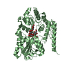
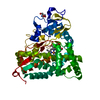
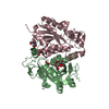
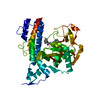
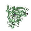
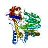
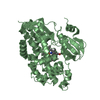
| Title | Partial Structure of the C-terminal domain of the HPIV4B phosphoprotein, fused to MBP. | |||||||||
 Components Components | Maltose-binding periplasmic protein, Phosphoprotein, chimeric construct | |||||||||
 Keywords Keywords | binding protein / viral protein / 3 helix bundle / viral nucleocapsid | |||||||||
| Function / homology |  Function and homology information Function and homology informationdetection of maltose stimulus / maltose transport complex / carbohydrate transport / carbohydrate transmembrane transporter activity / maltose binding / maltose transport / maltodextrin transmembrane transport / ATP-binding cassette (ABC) transporter complex, substrate-binding subunit-containing / ATP-binding cassette (ABC) transporter complex / cell chemotaxis ...detection of maltose stimulus / maltose transport complex / carbohydrate transport / carbohydrate transmembrane transporter activity / maltose binding / maltose transport / maltodextrin transmembrane transport / ATP-binding cassette (ABC) transporter complex, substrate-binding subunit-containing / ATP-binding cassette (ABC) transporter complex / cell chemotaxis / outer membrane-bounded periplasmic space / periplasmic space / DNA damage response / membrane Similarity search - Function | |||||||||
| Biological species |   Human parainfluenza virus 4b Human parainfluenza virus 4b | |||||||||
| Method |  X-RAY DIFFRACTION / MOLECULAR REPLACEMENT / Resolution: 2.21 Å X-RAY DIFFRACTION / MOLECULAR REPLACEMENT / Resolution: 2.21 Å | |||||||||
 Authors Authors | Yegambaram, K. / Bulloch, E.M.M. / Kingston, R.L. | |||||||||
 Citation Citation | Journal: Protein Sci. / Year: 2013 Title: Protein domain definition should allow for conditional disorder. Authors: Yegambaram, K. / Bulloch, E.M. / Kingston, R.L. | |||||||||
| History |
|
- Structure visualization
| Structure viewer | Molecule: MolmilJmol/JSmol |
|---|
- Downloads & links
-Download
| PDBx/mmCIF format | 4kyd.cif.gz | 327 KB | Display | PDBx/mmCIF format |
|---|---|---|---|---|
| PDB format | pdb4kyd.ent.gz | 268.1 KB | Display | PDB format |
| PDBx/mmJSON format | 4kyd.json.gz | Tree view | PDBx/mmJSON format | |
| Others |  Other downloads Other downloads |
-Validation report
| Arichive directory | https://data.pdbj.org/pub/pdb/validation_reports/ky/4kydftp://data.pdbj.org/pub/pdb/validation_reports/ky/4kyd | HTTPS FTP |
|---|
-Related structure data
| Related structure data |  4kycC  4kyeC 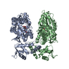 1hsjS C: citing same article ( S: Starting model for refinement |
|---|---|
| Similar structure data |


-Links
 PDBj
PDBj


- Assembly
| Deposited unit | 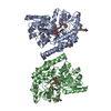
| ||||||||||||
|---|---|---|---|---|---|---|---|---|---|---|---|---|---|
| 1 | 
| ||||||||||||
| 2 | 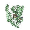
| ||||||||||||
| 3 |
| ||||||||||||
| Unit cell |
| ||||||||||||
| Components on special symmetry positions |
| ||||||||||||
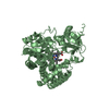
| Details | AUTHOR REMARK. IN SOLUTION THE C-TERMINAL DOMAIN OF THE HPIV4B PHOSPHOPROTEIN FORMS A MONOMERIC 3-HELIX BUNDLE. IN THE CRYSTAL, DISPLACEMENT OF THE FINAL HELIX AND PARTIAL REPACKING OF THE HYDROPHOBIC CORE LEAD TO FORMATION OF A HOMO-DIMERIC 4-HELIX BUNDLE. WE HAVE FOUND NO EVIDENCE THAT THIS HOMO-DIMER PERSISTS IN SOLUTION. SEE PRIMARY CITATION |
-Components
| #1: Protein | Mass: 45953.973 Da / Num. of mol.: 2 Fragment: unp P0AEX9 residues 27-392, unp P21738 residues 351-399 Mutation: D82A,K83A, E359A, K362A, D363A, C368S Source method: isolated from a genetically manipulated source Source: (gene. exp.) Human parainfluenza virus 4bStrain: K12, 68-333 Strain / Gene: b4034, JW3994, malE, P, P/V / Plasmid: pMAL(A) / Production host: #2: Polysaccharide |   Source method: isolated from a genetically manipulated source Details: oligosaccharide / References: alpha-maltose #3: Chemical | ChemComp-MPO /   Mass: 209.263 Da / Num. of mol.: 6 / Source method: obtained synthetically / Formula: C7H15NO4S / Comment: pH buffer*YM Mass: 209.263 Da / Num. of mol.: 6 / Source method: obtained synthetically / Formula: C7H15NO4S / Comment: pH buffer*YM#4: Water | ChemComp-HOH / |  Mass: 18.015 Da / Num. of mol.: 195 / Source method: isolated from a natural source / Formula: H2O Mass: 18.015 Da / Num. of mol.: 195 / Source method: isolated from a natural source / Formula: H2O |
|---|
-Experimental details
-Experiment
| Experiment | Method: X-RAY DIFFRACTION / Number of used crystals: 1 |
|---|
- Sample preparation
| Crystal | Density Matthews: 2.44 Å3/Da / Density % sol: 49.6 % |
|---|---|
| Crystal grow | Temperature: 277.15 K / Method: vapor diffusion, sitting drop / pH: 7.3 Details: 24 %(w/v) PEG 2000 0.2 M MOPS/KOH, pH 7.30, VAPOR DIFFUSION, SITTING DROP, temperature 277.15K |
-Data collection
| Diffraction | Mean temperature: 110 K |
|---|---|
| Diffraction source | Source: ROTATING ANODE / Type: RIGAKU MICROMAX-007 HF / Wavelength: 1.54179 Å |
| Detector | Type: MAR scanner 345 mm plate / Detector: IMAGE PLATE / Date: Sep 23, 2001 / Details: Osmic mirror optics |
| Radiation | Monochromator: Rigaku Varimax HF confocal / Protocol: SINGLE WAVELENGTH / Monochromatic (M) / Laue (L): M / Scattering type: x-ray |
| Radiation wavelength | Wavelength: 1.54179 Å / Relative weight: 1 |
| Reflection | Resolution: 2.21→38.1 Å / Num. all: 43158 / Num. obs: 43158 / % possible obs: 99.1 % / Observed criterion σ(F): 0 / Observed criterion σ(I): 0 / Redundancy: 9.5 % / Net I/σ(I): 4.3 |
| Reflection shell | Resolution: 2.21→2.25 Å / Redundancy: 6.9 % / Mean I/σ(I) obs: 1.7 / % possible all: 82.1 |
- Processing
| Software |
| ||||||||||||||||||||||||||||||||||||||||||||||||||||||||||||||||||||||||||||||||||||||||||||||||||||||||||||||||||||||||||||||||||||||||||||||||||||||||||||||||||||||||||||||||||||||
|---|---|---|---|---|---|---|---|---|---|---|---|---|---|---|---|---|---|---|---|---|---|---|---|---|---|---|---|---|---|---|---|---|---|---|---|---|---|---|---|---|---|---|---|---|---|---|---|---|---|---|---|---|---|---|---|---|---|---|---|---|---|---|---|---|---|---|---|---|---|---|---|---|---|---|---|---|---|---|---|---|---|---|---|---|---|---|---|---|---|---|---|---|---|---|---|---|---|---|---|---|---|---|---|---|---|---|---|---|---|---|---|---|---|---|---|---|---|---|---|---|---|---|---|---|---|---|---|---|---|---|---|---|---|---|---|---|---|---|---|---|---|---|---|---|---|---|---|---|---|---|---|---|---|---|---|---|---|---|---|---|---|---|---|---|---|---|---|---|---|---|---|---|---|---|---|---|---|---|---|---|---|---|---|
| Refinement | Method to determine structure: MOLECULAR REPLACEMENT Starting model: PDB accession code 1HSJ Resolution: 2.21→38.1 Å / Cor.coef. Fo:Fc: 0.965 / Cor.coef. Fo:Fc free: 0.945 / SU B: 10.274 / SU ML: 0.137 Isotropic thermal model: 1 TLS group per protein molecule + Residual Isotropic B-Factors Cross valid method: THROUGHOUT / ESU R: 0.259 / ESU R Free: 0.199 / Stereochemistry target values: MAXIMUM LIKELIHOOD
| ||||||||||||||||||||||||||||||||||||||||||||||||||||||||||||||||||||||||||||||||||||||||||||||||||||||||||||||||||||||||||||||||||||||||||||||||||||||||||||||||||||||||||||||||||||||
| Solvent computation | Ion probe radii: 0.8 Å / Shrinkage radii: 0.8 Å / VDW probe radii: 1.2 Å / Solvent model: MASK | ||||||||||||||||||||||||||||||||||||||||||||||||||||||||||||||||||||||||||||||||||||||||||||||||||||||||||||||||||||||||||||||||||||||||||||||||||||||||||||||||||||||||||||||||||||||
| Displacement parameters | Biso mean: 54.365 Å2
| ||||||||||||||||||||||||||||||||||||||||||||||||||||||||||||||||||||||||||||||||||||||||||||||||||||||||||||||||||||||||||||||||||||||||||||||||||||||||||||||||||||||||||||||||||||||
| Refinement step | Cycle: LAST / Resolution: 2.21→38.1 Å
| ||||||||||||||||||||||||||||||||||||||||||||||||||||||||||||||||||||||||||||||||||||||||||||||||||||||||||||||||||||||||||||||||||||||||||||||||||||||||||||||||||||||||||||||||||||||
| Refine LS restraints |
|