 Movie
Movie Controller

[English] 日本語
 Yorodumi
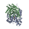
- PDB-6kfr: Amino-transferase (AMT) Domain - Arg Complex of Hybrid Polyketide... -
+ Open data

- Basic information
| Entry | Database: PDB / ID: 6kfr | ||||||
|---|---|---|---|---|---|---|---|
| Title | Amino-transferase (AMT) Domain - Arg Complex of Hybrid Polyketide/Non-Ribosomal Peptide Synthetase | ||||||
 Components Components | Mycosubtilin synthase subunit A | ||||||
 Keywords Keywords | TRANSFERASE / Amino-transferase / Hybrid Polyketide/Non-Ribosomal Peptide Synthetase | ||||||
| Function / homology |  Function and homology information Function and homology informationamino acid activation for nonribosomal peptide biosynthetic process / secondary metabolite biosynthetic process / transaminase activity / ligase activity / phosphopantetheine binding / 3-oxoacyl-[acyl-carrier-protein] synthase activity / antibiotic biosynthetic process / Transferases; Acyltransferases; Transferring groups other than aminoacyl groups / fatty acid biosynthetic process / pyridoxal phosphate binding / cytosol Similarity search - Function | ||||||
| Biological species |  | ||||||
| Method |  X-RAY DIFFRACTION / SYNCHROTRON / MOLECULAR REPLACEMENT / Resolution: 3.1 Å X-RAY DIFFRACTION / SYNCHROTRON / MOLECULAR REPLACEMENT / Resolution: 3.1 Å | ||||||
 Authors Authors | Tian, Q.W. / Jiang, T. | ||||||
| Funding support |  China, 1items China, 1items
| ||||||
 Citation Citation | Journal: To Be Published Title: Structure Insights into the Molecular Mechanism of Two Tailoring Domains of Hybrid Polyketide/Non-Ribosomal Peptide Synthetase Authors: Tian, Q.W. / Jiang, T. | ||||||
| History |
|
- Structure visualization
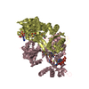
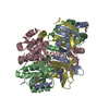
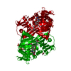
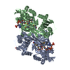
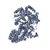
| Structure viewer | Molecule: MolmilJmol/JSmol |
|---|
- Downloads & links
-Download
| PDBx/mmCIF format | 6kfr.cif.gz | 185.9 KB | Display | PDBx/mmCIF format |
|---|---|---|---|---|
| PDB format | pdb6kfr.ent.gz | 145.6 KB | Display | PDB format |
| PDBx/mmJSON format | 6kfr.json.gz | Tree view | PDBx/mmJSON format | |
| Others |  Other downloads Other downloads |
-Validation report
| Arichive directory | https://data.pdbj.org/pub/pdb/validation_reports/kf/6kfrftp://data.pdbj.org/pub/pdb/validation_reports/kf/6kfr | HTTPS FTP |
|---|
-Related structure data
| Related structure data |  6ketC  6kfmSC  6kfuC S: Starting model for refinement C: citing same article ( |
|---|---|
| Similar structure data |


-Links
 PDBj
PDBj


- Assembly
| Deposited unit | 
| ||||||||||||
|---|---|---|---|---|---|---|---|---|---|---|---|---|---|
| 1 |
| ||||||||||||
| Unit cell |
|
-Components
| #1: Protein | Mass: 51454.594 Da / Num. of mol.: 2 Source method: isolated from a genetically manipulated source Details: SF file contains Friedel pairs. / Source: (gene. exp.) References: UniProt: Q9R9J1, Transferases; Acyltransferases; Transferring groups other than aminoacyl groups #2: Chemical |   Mass: 247.142 Da / Num. of mol.: 2 / Source method: obtained synthetically / Formula: C8H10NO6P / Feature type: SUBJECT OF INVESTIGATION Mass: 247.142 Da / Num. of mol.: 2 / Source method: obtained synthetically / Formula: C8H10NO6P / Feature type: SUBJECT OF INVESTIGATION#3: Chemical |   Type: L-peptide linking / Mass: 175.209 Da / Num. of mol.: 2 / Source method: obtained synthetically / Formula: C6H15N4O2 / Feature type: SUBJECT OF INVESTIGATION Type: L-peptide linking / Mass: 175.209 Da / Num. of mol.: 2 / Source method: obtained synthetically / Formula: C6H15N4O2 / Feature type: SUBJECT OF INVESTIGATIONHas ligand of interest | Y | Has protein modification | N | |
|---|
-Experimental details
-Experiment
| Experiment | Method: X-RAY DIFFRACTION / Number of used crystals: 1 |
|---|
- Sample preparation
| Crystal | Density Matthews: 2.3 Å3/Da / Density % sol: 46.58 % |
|---|---|
| Crystal grow | Temperature: 312 K / Method: vapor diffusion, hanging drop / pH: 6.7 Details: 10 mM MgCl2, 5 mM NiCl, 0.1 M HEPES, pH 6.7, 18% (v/v) PEG 3350 |
-Data collection
| Diffraction | Mean temperature: 100 K / Serial crystal experiment: N |
|---|---|
| Diffraction source | Source: SYNCHROTRON / Site: SSRF / Beamline: BL19U1 / Wavelength: 0.9793 Å |
| Detector | Type: DECTRIS PILATUS3 S 6M / Detector: PIXEL / Date: Jan 17, 2019 |
| Radiation | Protocol: SINGLE WAVELENGTH / Monochromatic (M) / Laue (L): M / Scattering type: x-ray |
| Radiation wavelength | Wavelength: 0.9793 Å / Relative weight: 1 |
| Reflection | Resolution: 3→50 Å / Num. obs: 18563 / % possible obs: 96.7 % / Redundancy: 11.7 % / CC1/2: 0.979 / Rpim(I) all: 0.064 / Rrim(I) all: 0.224 / Χ2: 1.024 / Net I/σ(I): 10.43 |
| Reflection shell | Resolution: 3→3.05 Å / Redundancy: 9.3 % / Mean I/σ(I) obs: 1.83 / Num. unique obs: 776 / CC1/2: 0.781 / Rpim(I) all: 0.279 / Rrim(I) all: 0.907 / Χ2: 0.916 / % possible all: 83.1 |
- Processing
| Software |
| ||||||||||||||||
|---|---|---|---|---|---|---|---|---|---|---|---|---|---|---|---|---|---|
| Refinement | Method to determine structure: MOLECULAR REPLACEMENT Starting model: 6KFM Resolution: 3.1→42.53 Å / Cross valid method: FREE R-VALUE
| ||||||||||||||||
| Displacement parameters | Biso mean: 45.29 Å2 | ||||||||||||||||
| Refinement step | Cycle: LAST / Resolution: 3.1→42.53 Å
| ||||||||||||||||
| LS refinement shell | Resolution: 3.1→3.211 Å
|
