 Movie
Movie Controller


+ Open data

- Basic information
| Entry | Database: PDB / ID: 5k8j | |||||||||
|---|---|---|---|---|---|---|---|---|---|---|
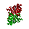
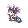
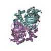
| Title | Structure of Caulobacter crescentus VapBC1 (apo form) | |||||||||
 Components Components |
| |||||||||
 Keywords Keywords | HYDROLASE / PIN domain / toxin-antitoxin / ribonuclease / DNA-binding | |||||||||
| Function / homology |  Function and homology information Function and homology informationRNA nuclease activity / toxin activity / Hydrolases; Acting on ester bonds / hydrolase activity / magnesium ion binding Similarity search - Function | |||||||||
| Biological species |  Caulobacter crescentus (bacteria) Caulobacter crescentus (bacteria) | |||||||||
| Method |  X-RAY DIFFRACTION / SYNCHROTRON / SAD / Resolution: 1.6 Å X-RAY DIFFRACTION / SYNCHROTRON / SAD / Resolution: 1.6 Å | |||||||||
 Authors Authors | Bendtsen, K.L. / Xu, K. / Luckmann, M. / Brodersen, D.E. | |||||||||
| Funding support |  Denmark, 2items Denmark, 2items
| |||||||||
 Citation Citation | Journal: Nucleic Acids Res. / Year: 2017 Title: Toxin inhibition in C. crescentus VapBC1 is mediated by a flexible pseudo-palindromic protein motif and modulated by DNA binding. Authors: Bendtsen, K.L. / Xu, K. / Luckmann, M. / Winther, K.S. / Shah, S.A. / Pedersen, C.N.S. / Brodersen, D.E. | |||||||||
| History |
|
- Structure visualization
| Structure viewer | Molecule: MolmilJmol/JSmol |
|---|
- Downloads & links
-Download
| PDBx/mmCIF format | 5k8j.cif.gz | 253.2 KB | Display | PDBx/mmCIF format |
|---|---|---|---|---|
| PDB format | pdb5k8j.ent.gz | 210.5 KB | Display | PDB format |
| PDBx/mmJSON format | 5k8j.json.gz | Tree view | PDBx/mmJSON format | |
| Others |  Other downloads Other downloads |
-Validation report
| Arichive directory | https://data.pdbj.org/pub/pdb/validation_reports/k8/5k8jftp://data.pdbj.org/pub/pdb/validation_reports/k8/5k8j | HTTPS FTP |
|---|
-Related structure data


-Links
 PDBj
PDBj

- Assembly
| Deposited unit | 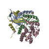
| ||||||||
|---|---|---|---|---|---|---|---|---|---|
| 1 | 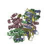
| ||||||||
| Unit cell |
| ||||||||
| Components on special symmetry positions |
|
-Components
| #1: Protein | Mass: 9548.433 Da / Num. of mol.: 2 Source method: isolated from a genetically manipulated source Source: (gene. exp.) Caulobacter crescentus (bacteria) / Strain: ATCC 19089 / CB15 / Gene: CC_0032 / Plasmid: pKW912HB / Production host: #2: Protein | Mass: 14288.886 Da / Num. of mol.: 2 Source method: isolated from a genetically manipulated source Source: (gene. exp.) Caulobacter crescentus (bacteria) / Strain: ATCC 19089 / CB15 / Gene: vapC, CC_0031 / Plasmid: pKW912HB / Production host: References: UniProt: Q9AC35, Hydrolases; Acting on ester bonds #3: Chemical |   Mass: 92.094 Da / Num. of mol.: 2 / Source method: obtained synthetically / Formula: C3H8O3 Mass: 92.094 Da / Num. of mol.: 2 / Source method: obtained synthetically / Formula: C3H8O3#4: Water | ChemComp-HOH / |  Mass: 18.015 Da / Num. of mol.: 352 / Source method: isolated from a natural source / Formula: H2O Mass: 18.015 Da / Num. of mol.: 352 / Source method: isolated from a natural source / Formula: H2OHas protein modification | Y | |
|---|
-Experimental details
-Experiment
| Experiment | Method: X-RAY DIFFRACTION / Number of used crystals: 1 |
|---|
- Sample preparation
| Crystal | Density Matthews: 2.57 Å3/Da / Density % sol: 52.17 % |
|---|---|
| Crystal grow | Temperature: 292.15 K / Method: vapor diffusion, sitting drop / pH: 5.5 Details: 3 M NaCl, 0.1 M Bis-tris pH 5.5, 5 mM MgCl2, 5 mM beta-mercapto ethanol, 25% glycerol |
-Data collection
| Diffraction | Mean temperature: 100 K |
|---|---|
| Diffraction source | Source: SYNCHROTRON / Site: MAX II  / Beamline: I911-3 / Wavelength: 0.972 Å / Beamline: I911-3 / Wavelength: 0.972 Å |
| Detector | Type: MARMOSAIC 225 mm CCD / Detector: CCD / Date: Apr 30, 2015 |
| Radiation | Protocol: SINGLE WAVELENGTH / Monochromatic (M) / Laue (L): M / Scattering type: x-ray |
| Radiation wavelength | Wavelength: 0.972 Å / Relative weight: 1 |
| Reflection | Resolution: 1.6→37.4 Å / Num. obs: 125126 / % possible obs: 100 % / Redundancy: 23.2 % / Biso Wilson estimate: 16.4 Å2 / CC1/2: 1 / Rsym value: 0.14 / Net I/σ(I): 22.8 |
| Reflection shell | Resolution: 1.6→1.64 Å / Redundancy: 22.8 % / Mean I/σ(I) obs: 1.7 / % possible all: 100 |
- Processing
| Software |
| |||||||||||||||||||||||||||||||||||||||||||||||||||||||||||||||||||||||||||||||||||||||||||||||||||||||||
|---|---|---|---|---|---|---|---|---|---|---|---|---|---|---|---|---|---|---|---|---|---|---|---|---|---|---|---|---|---|---|---|---|---|---|---|---|---|---|---|---|---|---|---|---|---|---|---|---|---|---|---|---|---|---|---|---|---|---|---|---|---|---|---|---|---|---|---|---|---|---|---|---|---|---|---|---|---|---|---|---|---|---|---|---|---|---|---|---|---|---|---|---|---|---|---|---|---|---|---|---|---|---|---|---|---|---|
| Refinement | Method to determine structure: SAD / Resolution: 1.6→37.4 Å / SU ML: 0.14 / Cross valid method: FREE R-VALUE / σ(F): 0 / Phase error: 16.65
| |||||||||||||||||||||||||||||||||||||||||||||||||||||||||||||||||||||||||||||||||||||||||||||||||||||||||
| Solvent computation | Shrinkage radii: 0.9 Å / VDW probe radii: 1.11 Å | |||||||||||||||||||||||||||||||||||||||||||||||||||||||||||||||||||||||||||||||||||||||||||||||||||||||||
| Displacement parameters | Biso mean: 27.21 Å2 | |||||||||||||||||||||||||||||||||||||||||||||||||||||||||||||||||||||||||||||||||||||||||||||||||||||||||
| Refinement step | Cycle: LAST / Resolution: 1.6→37.4 Å
| |||||||||||||||||||||||||||||||||||||||||||||||||||||||||||||||||||||||||||||||||||||||||||||||||||||||||
| Refine LS restraints |
| |||||||||||||||||||||||||||||||||||||||||||||||||||||||||||||||||||||||||||||||||||||||||||||||||||||||||
| LS refinement shell |
| |||||||||||||||||||||||||||||||||||||||||||||||||||||||||||||||||||||||||||||||||||||||||||||||||||||||||
| Refinement TLS params. | Method: refined / Origin x: 20.3497 Å / Origin y: 36.3746 Å / Origin z: 39.8612 Å
| |||||||||||||||||||||||||||||||||||||||||||||||||||||||||||||||||||||||||||||||||||||||||||||||||||||||||
| Refinement TLS group | Selection details: all |
