 Movie
Movie Controller

[English] 日本語
 Yorodumi
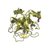
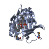
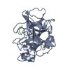
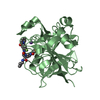
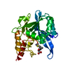
- PDB-6kds: Crystal structure of human NRMT2 in complex with alpha-N-monometh... -
+ Open data

- Basic information
| Entry | Database: PDB / ID: 6kds | ||||||
|---|---|---|---|---|---|---|---|
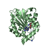
| Title | Crystal structure of human NRMT2 in complex with alpha-N-monomethylated human CENP-A peptide | ||||||
 Components Components |
| ||||||
 Keywords Keywords | TRANSFERASE / core methyltransferase fold | ||||||
| Function / homology |  Function and homology information Function and homology informationprotein N-terminal monomethyltransferase / N-terminal protein amino acid methylation / N-terminal protein N-methyltransferase activity / CENP-A containing chromatin assembly / protein localization to chromosome, centromeric region / kinetochore assembly / condensed chromosome, centromeric region / establishment of mitotic spindle orientation / mitotic cytokinesis / chromosome, centromeric region ...protein N-terminal monomethyltransferase / N-terminal protein amino acid methylation / N-terminal protein N-methyltransferase activity / CENP-A containing chromatin assembly / protein localization to chromosome, centromeric region / kinetochore assembly / condensed chromosome, centromeric region / establishment of mitotic spindle orientation / mitotic cytokinesis / chromosome, centromeric region / pericentric heterochromatin / protein localization to CENP-A containing chromatin / CENP-A containing nucleosome / Amplification of signal from unattached kinetochores via a MAD2 inhibitory signal / Deposition of new CENPA-containing nucleosomes at the centromere / Mitotic Prometaphase / EML4 and NUDC in mitotic spindle formation / Resolution of Sister Chromatid Cohesion / RHO GTPases Activate Formins / structural constituent of chromatin / Separation of Sister Chromatids / nucleosome / protein heterodimerization activity / chromatin binding / DNA binding / nucleoplasm / nucleus / cytoplasm / cytosol Similarity search - Function | ||||||
| Biological species |  Homo sapiens (human) Homo sapiens (human) | ||||||
| Method |  X-RAY DIFFRACTION / SYNCHROTRON / MOLECULAR REPLACEMENT / Resolution: 1.844 Å X-RAY DIFFRACTION / SYNCHROTRON / MOLECULAR REPLACEMENT / Resolution: 1.844 Å | ||||||
 Authors Authors | Yue, Y. / Li, H. | ||||||
 Citation Citation | Journal: To be published Title: Substrate engagement regulates state-specific alpha-N methylation of CENP-A by NRMT2 Authors: Wu, R. / Yue, Y. / Zheng, X. / Li, H. | ||||||
| History |
|
- Structure visualization
| Structure viewer | Molecule: MolmilJmol/JSmol |
|---|
- Downloads & links
-Download
| PDBx/mmCIF format | 6kds.cif.gz | 116.4 KB | Display | PDBx/mmCIF format |
|---|---|---|---|---|
| PDB format | pdb6kds.ent.gz | 86.6 KB | Display | PDB format |
| PDBx/mmJSON format | 6kds.json.gz | Tree view | PDBx/mmJSON format | |
| Others |  Other downloads Other downloads |
-Validation report
| Arichive directory | https://data.pdbj.org/pub/pdb/validation_reports/kd/6kdsftp://data.pdbj.org/pub/pdb/validation_reports/kd/6kds | HTTPS FTP |
|---|
-Related structure data
| Related structure data |  6kdqC  5cvdS S: Starting model for refinement C: citing same article ( |
|---|---|
| Similar structure data |


-Links
 PDBj
PDBj


- Assembly
| Deposited unit | 
| ||||||||
|---|---|---|---|---|---|---|---|---|---|
| 1 |
| ||||||||
| Unit cell |
|
-Components
| #1: Protein | Mass: 27519.480 Da / Num. of mol.: 1 Source method: isolated from a genetically manipulated source Source: (gene. exp.) Homo sapiens (human) / Gene: METTL11B, C1orf184, NRMT2 / Plasmid: pET28 / Production host:  References: UniProt: Q5VVY1, protein N-terminal monomethyltransferase |
|---|---|
| #2: Protein/peptide | Mass: 744.867 Da / Num. of mol.: 1 / Source method: obtained synthetically / Source: (synth.) Homo sapiens (human) / References: UniProt: P49450*PLUS |
| #3: Chemical | ChemComp-SAH /   Mass: 384.411 Da / Num. of mol.: 1 / Source method: obtained synthetically / Formula: C14H20N6O5S Mass: 384.411 Da / Num. of mol.: 1 / Source method: obtained synthetically / Formula: C14H20N6O5S |
| #4: Water | ChemComp-HOH /  Mass: 18.015 Da / Num. of mol.: 167 / Source method: isolated from a natural source / Formula: H2O Mass: 18.015 Da / Num. of mol.: 167 / Source method: isolated from a natural source / Formula: H2O |
| Has ligand of interest | Y |
| Has protein modification | Y |
-Experimental details
-Experiment
| Experiment | Method: X-RAY DIFFRACTION / Number of used crystals: 1 |
|---|
- Sample preparation
| Crystal | Density Matthews: 2.32 Å3/Da / Density % sol: 47.08 % / Mosaicity: 0.886 ° / Mosaicity esd: 0.007 ° |
|---|---|
| Crystal grow | Temperature: 291 K / Method: vapor diffusion / pH: 8.1 / Details: 0.1M Tris pH 8.1, 14% w/v Polyethylene glycol 8000 |
-Data collection
| Diffraction | Mean temperature: 100 K / Serial crystal experiment: N | |||||||||||||||||||||||||||||||||||||||||||||||||||||||||||||||||||||||||||||||||||||||||||||||||||||||||||||||||||||||||||||||||||||||||||||||||||||||||||||||||||||||||||||||||||||||||||||
|---|---|---|---|---|---|---|---|---|---|---|---|---|---|---|---|---|---|---|---|---|---|---|---|---|---|---|---|---|---|---|---|---|---|---|---|---|---|---|---|---|---|---|---|---|---|---|---|---|---|---|---|---|---|---|---|---|---|---|---|---|---|---|---|---|---|---|---|---|---|---|---|---|---|---|---|---|---|---|---|---|---|---|---|---|---|---|---|---|---|---|---|---|---|---|---|---|---|---|---|---|---|---|---|---|---|---|---|---|---|---|---|---|---|---|---|---|---|---|---|---|---|---|---|---|---|---|---|---|---|---|---|---|---|---|---|---|---|---|---|---|---|---|---|---|---|---|---|---|---|---|---|---|---|---|---|---|---|---|---|---|---|---|---|---|---|---|---|---|---|---|---|---|---|---|---|---|---|---|---|---|---|---|---|---|---|---|---|---|---|---|
| Diffraction source | Source: SYNCHROTRON / Site: SSRF  / Beamline: BL17U / Wavelength: 0.97915 Å / Beamline: BL17U / Wavelength: 0.97915 Å | |||||||||||||||||||||||||||||||||||||||||||||||||||||||||||||||||||||||||||||||||||||||||||||||||||||||||||||||||||||||||||||||||||||||||||||||||||||||||||||||||||||||||||||||||||||||||||||
| Detector | Type: ADSC QUANTUM 315r / Detector: CCD / Date: May 21, 2014 / Details: mirrors | |||||||||||||||||||||||||||||||||||||||||||||||||||||||||||||||||||||||||||||||||||||||||||||||||||||||||||||||||||||||||||||||||||||||||||||||||||||||||||||||||||||||||||||||||||||||||||||
| Radiation | Monochromator: double crystal, Si(111) / Protocol: SINGLE WAVELENGTH / Monochromatic (M) / Laue (L): M / Scattering type: x-ray | |||||||||||||||||||||||||||||||||||||||||||||||||||||||||||||||||||||||||||||||||||||||||||||||||||||||||||||||||||||||||||||||||||||||||||||||||||||||||||||||||||||||||||||||||||||||||||||
| Radiation wavelength | Wavelength: 0.97915 Å / Relative weight: 1 | |||||||||||||||||||||||||||||||||||||||||||||||||||||||||||||||||||||||||||||||||||||||||||||||||||||||||||||||||||||||||||||||||||||||||||||||||||||||||||||||||||||||||||||||||||||||||||||
| Reflection | Resolution: 1.8→50 Å / Num. obs: 47754 / % possible obs: 98.1 % / Redundancy: 3.2 % / Rmerge(I) obs: 0.092 / Rpim(I) all: 0.06 / Rrim(I) all: 0.107 / Χ2: 2.722 / Net I/σ(I): 10.8 / Num. measured all: 154647 | |||||||||||||||||||||||||||||||||||||||||||||||||||||||||||||||||||||||||||||||||||||||||||||||||||||||||||||||||||||||||||||||||||||||||||||||||||||||||||||||||||||||||||||||||||||||||||||
| Reflection shell | Diffraction-ID: 1
|
- Processing
| Software |
| ||||||||||||||||||||||||||||||||||||||||||||||||||||||
|---|---|---|---|---|---|---|---|---|---|---|---|---|---|---|---|---|---|---|---|---|---|---|---|---|---|---|---|---|---|---|---|---|---|---|---|---|---|---|---|---|---|---|---|---|---|---|---|---|---|---|---|---|---|---|---|
| Refinement | Method to determine structure: MOLECULAR REPLACEMENT Starting model: 5CVD Resolution: 1.844→32.979 Å / SU ML: 0.18 / Cross valid method: THROUGHOUT / σ(F): 1.36 / Phase error: 21.04
| ||||||||||||||||||||||||||||||||||||||||||||||||||||||
| Solvent computation | Shrinkage radii: 0.9 Å / VDW probe radii: 1.11 Å | ||||||||||||||||||||||||||||||||||||||||||||||||||||||
| Displacement parameters | Biso max: 96.3 Å2 / Biso mean: 31.1282 Å2 / Biso min: 12.25 Å2 | ||||||||||||||||||||||||||||||||||||||||||||||||||||||
| Refinement step | Cycle: final / Resolution: 1.844→32.979 Å
| ||||||||||||||||||||||||||||||||||||||||||||||||||||||
| LS refinement shell | Refine-ID: X-RAY DIFFRACTION / Rfactor Rfree error: 0
| ||||||||||||||||||||||||||||||||||||||||||||||||||||||
| Refinement TLS params. | Method: refined / Origin x: 20.7037 Å / Origin y: 17.6994 Å / Origin z: 17.7445 Å
| ||||||||||||||||||||||||||||||||||||||||||||||||||||||
| Refinement TLS group |
|
