 Movie
Movie Controller

[English] 日本語
 Yorodumi
- PDB-6k52: Hyperthermophilic GH6 cellobiohydrolase (HmCel6A) from the microb... -
+ Open data

- Basic information
| Entry | Database: PDB / ID: 6k52 | ||||||
|---|---|---|---|---|---|---|---|
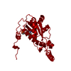
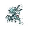
| Title | Hyperthermophilic GH6 cellobiohydrolase (HmCel6A) from the microbial flora of a Japanese hot spring | ||||||
 Components Components | GH6 cellobiohydrolase, HMCEL6A | ||||||
 Keywords Keywords | HYDROLASE / glycoside hydrolase / GH6 / thermostable | ||||||
| Function / homology | 1, 4-beta cellobiohydrolase / TIM Barrel / Alpha-Beta Barrel / Alpha Beta / ACETATE ION Function and homology information Function and homology information | ||||||
| Biological species | metagenome (others) | ||||||
| Method |  X-RAY DIFFRACTION / SYNCHROTRON / MOLECULAR REPLACEMENT / Resolution: 1.68000139531 Å X-RAY DIFFRACTION / SYNCHROTRON / MOLECULAR REPLACEMENT / Resolution: 1.68000139531 Å | ||||||
 Authors Authors | Baba, S. / Takeda, M. / Okuma, J. / Hirose, Y. / Nishimura, A. / Takata, M. / Oda, K. / Shibata, D. / Kondo, Y. / Kumasaka, T. | ||||||
 Citation Citation | Journal: To Be Published Title: A hyperthermophilic cellobiohydrolase mined from a hot spring metagenomic data Authors: Takeda, M. / Baba, S. / Okuma, J. / Hirose, Y. / Nishimura, A. / Takata, M. / Oda, K. / Shibata, D. / Kondo, Y. / Kumasaka, T. | ||||||
| History |
|
- Structure visualization
| Structure viewer | Molecule: MolmilJmol/JSmol |
|---|
- Downloads & links
-Download
| PDBx/mmCIF format | 6k52.cif.gz | 130.7 KB | Display | PDBx/mmCIF format |
|---|---|---|---|---|
| PDB format | pdb6k52.ent.gz | 86.9 KB | Display | PDB format |
| PDBx/mmJSON format | 6k52.json.gz | Tree view | PDBx/mmJSON format | |
| Others |  Other downloads Other downloads |
-Validation report
| Arichive directory | https://data.pdbj.org/pub/pdb/validation_reports/k5/6k52ftp://data.pdbj.org/pub/pdb/validation_reports/k5/6k52 | HTTPS FTP |
|---|
-Related structure data
| Related structure data |  6k55C  1oc5S S: Starting model for refinement C: citing same article ( |
|---|---|
| Similar structure data |


-Links
 PDBj
PDBj- Assembly
| Deposited unit | 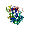
| ||||||||||||
|---|---|---|---|---|---|---|---|---|---|---|---|---|---|
| 1 |
| ||||||||||||
| Unit cell |
| ||||||||||||
| Components on special symmetry positions |
|
-Components
| #1: Protein | Mass: 46898.242 Da / Num. of mol.: 1 Source method: isolated from a genetically manipulated source Source: (gene. exp.) metagenome (others) / Production host:  References: cellulose 1,4-beta-cellobiosidase (non-reducing end) | ||||||||
|---|---|---|---|---|---|---|---|---|---|
| #2: Chemical |   Mass: 40.078 Da / Num. of mol.: 3 / Source method: obtained synthetically / Formula: Ca / Feature type: SUBJECT OF INVESTIGATION Mass: 40.078 Da / Num. of mol.: 3 / Source method: obtained synthetically / Formula: Ca / Feature type: SUBJECT OF INVESTIGATION#3: Chemical | ChemComp-ACT / |   Mass: 59.044 Da / Num. of mol.: 1 / Source method: obtained synthetically / Formula: C2H3O2 Mass: 59.044 Da / Num. of mol.: 1 / Source method: obtained synthetically / Formula: C2H3O2#4: Water | ChemComp-HOH / |  Mass: 18.015 Da / Num. of mol.: 589 / Source method: isolated from a natural source / Formula: H2O Mass: 18.015 Da / Num. of mol.: 589 / Source method: isolated from a natural source / Formula: H2OHas protein modification | Y | Sequence details | FIRST MET IS A INITIATING METHIONINE. THIS DNA SEQUENCE HAS BEEN DEPOSITED TO DDBJ WITH A ...FIRST MET IS A INITIATING | |
-Experimental details
-Experiment
| Experiment | Method: X-RAY DIFFRACTION / Number of used crystals: 1 |
|---|
- Sample preparation
| Crystal | Density Matthews: 4.56 Å3/Da / Density % sol: 73.05 % |
|---|---|
| Crystal grow | Temperature: 293 K / Method: vapor diffusion, sitting drop / pH: 8 Details: 20% (W/V) PEG 1000, 0.2M CAICIUM ACETATE, 0.1M IMIDAZOLE (PH 8.0) |
-Data collection
| Diffraction | Mean temperature: 100 K / Serial crystal experiment: N |
|---|---|
| Diffraction source | Source: SYNCHROTRON / Site: SPring-8  / Beamline: BL38B1 / Wavelength: 1 Å / Beamline: BL38B1 / Wavelength: 1 Å |
| Detector | Type: ADSC QUANTUM 315r / Detector: CCD / Date: Oct 29, 2012 |
| Radiation | Monochromator: SI (111) / Protocol: SINGLE WAVELENGTH / Monochromatic (M) / Laue (L): M / Scattering type: x-ray |
| Radiation wavelength | Wavelength: 1 Å / Relative weight: 1 |
| Reflection | Resolution: 1.68→50 Å / Num. obs: 97033 / % possible obs: 100 % / Redundancy: 16.3 % / Biso Wilson estimate: 17.3900593193 Å2 / CC1/2: 1 / Rmerge(I) obs: 0.071 / Rpim(I) all: 0.018 / Rrim(I) all: 0.074 / Net I/σ(I): 36.7 |
| Reflection shell | Resolution: 1.68→1.74 Å / Redundancy: 15.7 % / Rmerge(I) obs: 0.597 / Mean I/σ(I) obs: 6.6 / Num. unique obs: 9626 / CC1/2: 0.961 / Rpim(I) all: 0.155 / Rrim(I) all: 0.617 / % possible all: 100 |
- Processing
| Software |
| |||||||||||||||||||||||||||||||||||||||||||||||||||||||||||||||||||||||||||||||||||||||||||||||||||||||||||||||||||||||||||||||||||||||||||||||||||||||||||||||||||||||||||||||||||||||||||||||||||||||||||||||||||||||||
|---|---|---|---|---|---|---|---|---|---|---|---|---|---|---|---|---|---|---|---|---|---|---|---|---|---|---|---|---|---|---|---|---|---|---|---|---|---|---|---|---|---|---|---|---|---|---|---|---|---|---|---|---|---|---|---|---|---|---|---|---|---|---|---|---|---|---|---|---|---|---|---|---|---|---|---|---|---|---|---|---|---|---|---|---|---|---|---|---|---|---|---|---|---|---|---|---|---|---|---|---|---|---|---|---|---|---|---|---|---|---|---|---|---|---|---|---|---|---|---|---|---|---|---|---|---|---|---|---|---|---|---|---|---|---|---|---|---|---|---|---|---|---|---|---|---|---|---|---|---|---|---|---|---|---|---|---|---|---|---|---|---|---|---|---|---|---|---|---|---|---|---|---|---|---|---|---|---|---|---|---|---|---|---|---|---|---|---|---|---|---|---|---|---|---|---|---|---|---|---|---|---|---|---|---|---|---|---|---|---|---|---|---|---|---|---|---|---|---|
| Refinement | Method to determine structure: MOLECULAR REPLACEMENT Starting model: 1OC5 Resolution: 1.68000139531→32.9103974351 Å / SU ML: 0.176042297081 / Cross valid method: FREE R-VALUE / σ(F): 1.33476296841 / Phase error: 21.1813420271
| |||||||||||||||||||||||||||||||||||||||||||||||||||||||||||||||||||||||||||||||||||||||||||||||||||||||||||||||||||||||||||||||||||||||||||||||||||||||||||||||||||||||||||||||||||||||||||||||||||||||||||||||||||||||||
| Solvent computation | Shrinkage radii: 0.9 Å / VDW probe radii: 1.11 Å | |||||||||||||||||||||||||||||||||||||||||||||||||||||||||||||||||||||||||||||||||||||||||||||||||||||||||||||||||||||||||||||||||||||||||||||||||||||||||||||||||||||||||||||||||||||||||||||||||||||||||||||||||||||||||
| Displacement parameters | Biso mean: 21.3657637173 Å2 | |||||||||||||||||||||||||||||||||||||||||||||||||||||||||||||||||||||||||||||||||||||||||||||||||||||||||||||||||||||||||||||||||||||||||||||||||||||||||||||||||||||||||||||||||||||||||||||||||||||||||||||||||||||||||
| Refinement step | Cycle: LAST / Resolution: 1.68000139531→32.9103974351 Å
| |||||||||||||||||||||||||||||||||||||||||||||||||||||||||||||||||||||||||||||||||||||||||||||||||||||||||||||||||||||||||||||||||||||||||||||||||||||||||||||||||||||||||||||||||||||||||||||||||||||||||||||||||||||||||
| Refine LS restraints |
| |||||||||||||||||||||||||||||||||||||||||||||||||||||||||||||||||||||||||||||||||||||||||||||||||||||||||||||||||||||||||||||||||||||||||||||||||||||||||||||||||||||||||||||||||||||||||||||||||||||||||||||||||||||||||
| LS refinement shell |
|
