 Movie
Movie Controller


+ Open data

- Basic information
| Entry | Database: PDB / ID: 1nox | ||||||
|---|---|---|---|---|---|---|---|
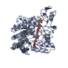
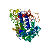
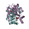
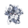
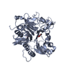
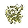
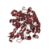
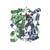
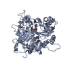
| Title | NADH OXIDASE FROM THERMUS THERMOPHILUS | ||||||
 Components Components | NADH OXIDASE | ||||||
 Keywords Keywords | FLAVOENZYME / FLAVOPROTEIN FMN / OXIDOREDUCTASE / THERMOPHILE | ||||||
| Function / homology |  Function and homology information Function and homology informationNADH:ubiquinone reductase (H+-translocating) / NADH dehydrogenase (ubiquinone) activity / oxidoreductase activity Similarity search - Function | ||||||
| Biological species |   Thermus thermophilus (bacteria) Thermus thermophilus (bacteria) | ||||||
| Method |  X-RAY DIFFRACTION / SYNCHROTRON / ISOMORPHOUS, MOLECULAR REPLACEMENT / Resolution: 1.59 Å X-RAY DIFFRACTION / SYNCHROTRON / ISOMORPHOUS, MOLECULAR REPLACEMENT / Resolution: 1.59 Å | ||||||
 Authors Authors | Hecht, H.J. / Erdmann, H. / Park, H.J. / Sprinzl, M. / Schmid, R.D. | ||||||
 Citation Citation | Journal: Nat.Struct.Biol. / Year: 1995 Title: Crystal structure of NADH oxidase from Thermus thermophilus. Authors: Hecht, H.J. / Erdmann, H. / Park, H.J. / Sprinzl, M. / Schmid, R.D. | ||||||
| History |
|
- Structure visualization
| Structure viewer | Molecule: MolmilJmol/JSmol |
|---|
- Downloads & links
-Download
| PDBx/mmCIF format | 1nox.cif.gz | 54.3 KB | Display | PDBx/mmCIF format |
|---|---|---|---|---|
| PDB format | pdb1nox.ent.gz | 38.6 KB | Display | PDB format |
| PDBx/mmJSON format | 1nox.json.gz | Tree view | PDBx/mmJSON format | |
| Others |  Other downloads Other downloads |
-Validation report
| Arichive directory | https://data.pdbj.org/pub/pdb/validation_reports/no/1noxftp://data.pdbj.org/pub/pdb/validation_reports/no/1nox | HTTPS FTP |
|---|
-Related structure data
| Similar structure data |
|---|

-Links
 PDBj
PDBj- Assembly
| Deposited unit | 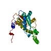
| ||||||||
|---|---|---|---|---|---|---|---|---|---|
| 1 | 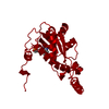
| ||||||||
| Unit cell |
|
-Components
| #1: Protein | Mass: 22780.455 Da / Num. of mol.: 1 Source method: isolated from a genetically manipulated source Source: (gene. exp.) Thermus thermophilus (bacteria) / Strain: HB8 / Plasmid: PTNADOX / Production host: |
|---|---|
| #2: Chemical | ChemComp-FMN /   Mass: 456.344 Da / Num. of mol.: 1 / Source method: obtained synthetically / Formula: C17H21N4O9P Mass: 456.344 Da / Num. of mol.: 1 / Source method: obtained synthetically / Formula: C17H21N4O9P |
| #3: Water | ChemComp-HOH /  Mass: 18.015 Da / Num. of mol.: 93 / Source method: isolated from a natural source / Formula: H2O Mass: 18.015 Da / Num. of mol.: 93 / Source method: isolated from a natural source / Formula: H2O |
-Experimental details
-Experiment
| Experiment | Method: X-RAY DIFFRACTION / Number of used crystals: 1 |
|---|
- Sample preparation
| Crystal | Density Matthews: 2.4 Å3/Da / Density % sol: 47 % | ||||||||||||||||||||||||||||||||||||
|---|---|---|---|---|---|---|---|---|---|---|---|---|---|---|---|---|---|---|---|---|---|---|---|---|---|---|---|---|---|---|---|---|---|---|---|---|---|
| Crystal grow | pH: 6 Details: 0.05 M MES-TRIS BUFFER PH 6.0, 26 % PEG 4000, 0.3 M NACL, 50 MM FMN | ||||||||||||||||||||||||||||||||||||
| Crystal grow | *PLUS Temperature: 12 ℃ / pH: 6.6 / Method: vapor diffusion, sitting drop | ||||||||||||||||||||||||||||||||||||
| Components of the solutions | *PLUS
|
-Data collection
| Diffraction | Mean temperature: 283 K |
|---|---|
| Diffraction source | Source: SYNCHROTRON / Site: MPG/DESY, HAMBURG  / Beamline: BW6 / Wavelength: 1 / Beamline: BW6 / Wavelength: 1 |
| Detector | Type: MARRESEARCH / Detector: IMAGE PLATE / Date: Nov 1, 1993 / Details: MIRRORS |
| Radiation | Monochromator: SI(111) / Monochromatic (M) / Laue (L): M / Scattering type: x-ray |
| Radiation wavelength | Wavelength: 1 Å / Relative weight: 1 |
| Reflection | Highest resolution: 1.59 Å / Num. obs: 28064 / % possible obs: 96.2 % / Observed criterion σ(I): 0 / Redundancy: 3.9 % / Biso Wilson estimate: 18.2 Å2 / Rmerge(I) obs: 0.071 / Rsym value: 0.069 / Net I/σ(I): 6.2 |
| Reflection shell | Resolution: 1.59→1.68 Å / Redundancy: 3.8 % / Rmerge(I) obs: 0.071 / Mean I/σ(I) obs: 3.2 / Rsym value: 0.071 / % possible all: 95.3 |
| Reflection shell | *PLUS % possible obs: 95.3 % |
- Processing
| Software |
| ||||||||||||||||||||||||||||||||||||||||||||||||||||||||||||||||||||||||||||||||||||
|---|---|---|---|---|---|---|---|---|---|---|---|---|---|---|---|---|---|---|---|---|---|---|---|---|---|---|---|---|---|---|---|---|---|---|---|---|---|---|---|---|---|---|---|---|---|---|---|---|---|---|---|---|---|---|---|---|---|---|---|---|---|---|---|---|---|---|---|---|---|---|---|---|---|---|---|---|---|---|---|---|---|---|---|---|---|
| Refinement | Method to determine structure: ISOMORPHOUS, MOLECULAR REPLACEMENT Starting model: MODEL FROM ISOMORPHOUS REPLACEMENT Resolution: 1.59→21 Å / σ(F): 0 / Details: FREE-R-VALUE INTRODUCED LATE IN REFINEMENT.
| ||||||||||||||||||||||||||||||||||||||||||||||||||||||||||||||||||||||||||||||||||||
| Displacement parameters | Biso mean: 23.3 Å2 | ||||||||||||||||||||||||||||||||||||||||||||||||||||||||||||||||||||||||||||||||||||
| Refine analyze | Luzzati d res low obs: 7 Å / Luzzati sigma a obs: 0.03 Å | ||||||||||||||||||||||||||||||||||||||||||||||||||||||||||||||||||||||||||||||||||||
| Refinement step | Cycle: LAST / Resolution: 1.59→21 Å
| ||||||||||||||||||||||||||||||||||||||||||||||||||||||||||||||||||||||||||||||||||||
| Refine LS restraints |
| ||||||||||||||||||||||||||||||||||||||||||||||||||||||||||||||||||||||||||||||||||||
| Software | *PLUS Name: REFMAC / Classification: refinement | ||||||||||||||||||||||||||||||||||||||||||||||||||||||||||||||||||||||||||||||||||||
| Refinement | *PLUS | ||||||||||||||||||||||||||||||||||||||||||||||||||||||||||||||||||||||||||||||||||||
| Solvent computation | *PLUS | ||||||||||||||||||||||||||||||||||||||||||||||||||||||||||||||||||||||||||||||||||||
| Displacement parameters | *PLUS |
