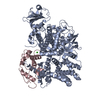
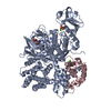
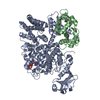
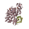
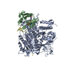
[English] 日本語
Yorodumi
- PDB-6k4r: Crystal structure of SidJ-CaM-AMP ternary complex at 3.11 A -

+
Open data


ID or keywords:

Loading...

-
Basic information

Entry
Database: PDB / ID: 6k4r
TitleCrystal structure of SidJ-CaM-AMP ternary complex at 3.11 A
Components
  • Calmodulin-1
  • SidJ
KeywordsTRANSFERASE/CELL CYCLE / A novel kinase / TRANSFERASE / TRANSFERASE-CELL CYCLE complex
Function / homology
Function and homology information


protein-glutamic acid ligase activity, initiating / protein-glutamic acid ligase activity, elongating / Ligases / CaM pathway / Cam-PDE 1 activation / Sodium/Calcium exchangers / Calmodulin induced events / positive regulation of ryanodine-sensitive calcium-release channel activity / Reduction of cytosolic Ca++ levels / Activation of Ca-permeable Kainate Receptor ...protein-glutamic acid ligase activity, initiating / protein-glutamic acid ligase activity, elongating / Ligases / CaM pathway / Cam-PDE 1 activation / Sodium/Calcium exchangers / Calmodulin induced events / positive regulation of ryanodine-sensitive calcium-release channel activity / Reduction of cytosolic Ca++ levels / Activation of Ca-permeable Kainate Receptor / CREB1 phosphorylation through the activation of CaMKII/CaMKK/CaMKIV cascasde / Loss of phosphorylation of MECP2 at T308 / CREB1 phosphorylation through the activation of Adenylate Cyclase / CaMK IV-mediated phosphorylation of CREB / PKA activation / negative regulation of high voltage-gated calcium channel activity / Glycogen breakdown (glycogenolysis) / CLEC7A (Dectin-1) induces NFAT activation / Activation of RAC1 downstream of NMDARs / organelle localization by membrane tethering / negative regulation of ryanodine-sensitive calcium-release channel activity / mitochondrion-endoplasmic reticulum membrane tethering / autophagosome membrane docking / negative regulation of calcium ion export across plasma membrane / regulation of cardiac muscle cell action potential / presynaptic endocytosis / Synthesis of IP3 and IP4 in the cytosol / regulation of cell communication by electrical coupling involved in cardiac conduction / Phase 0 - rapid depolarisation / calcineurin-mediated signaling / Negative regulation of NMDA receptor-mediated neuronal transmission / Unblocking of NMDA receptors, glutamate binding and activation / RHO GTPases activate PAKs / Ion transport by P-type ATPases / Uptake and function of anthrax toxins / regulation of ryanodine-sensitive calcium-release channel activity / Long-term potentiation / protein phosphatase activator activity / Calcineurin activates NFAT / Regulation of MECP2 expression and activity / DARPP-32 events / catalytic complex / Smooth Muscle Contraction / detection of calcium ion / regulation of cardiac muscle contraction / RHO GTPases activate IQGAPs / regulation of cardiac muscle contraction by regulation of the release of sequestered calcium ion / calcium channel inhibitor activity / cellular response to interferon-beta / Protein methylation / presynaptic cytosol / Activation of AMPK downstream of NMDARs / Ion homeostasis / regulation of release of sequestered calcium ion into cytosol by sarcoplasmic reticulum / eNOS activation / titin binding / Tetrahydrobiopterin (BH4) synthesis, recycling, salvage and regulation / sperm midpiece / voltage-gated potassium channel complex / regulation of calcium-mediated signaling / calcium channel complex / substantia nigra development / FCERI mediated Ca+2 mobilization / cysteine-type peptidase activity / Ras activation upon Ca2+ influx through NMDA receptor / regulation of heart rate / FCGR3A-mediated IL10 synthesis / calyx of Held / Antigen activates B Cell Receptor (BCR) leading to generation of second messengers / adenylate cyclase activator activity / sarcomere / VEGFR2 mediated cell proliferation / regulation of cytokinesis / protein serine/threonine kinase activator activity / VEGFR2 mediated vascular permeability / spindle microtubule / Translocation of SLC2A4 (GLUT4) to the plasma membrane / positive regulation of receptor signaling pathway via JAK-STAT / RAF activation / Transcriptional activation of mitochondrial biogenesis / cellular response to type II interferon / Stimuli-sensing channels / long-term synaptic potentiation / response to calcium ion / spindle pole / RAS processing / Signaling by RAF1 mutants / calcium-dependent protein binding / Signaling by moderate kinase activity BRAF mutants / Paradoxical activation of RAF signaling by kinase inactive BRAF / Signaling downstream of RAS mutants / G2/M transition of mitotic cell cycle / Signaling by BRAF and RAF1 fusions / Inactivation, recovery and regulation of the phototransduction cascade / Platelet degranulation / myelin sheath / transferase activity / Ca2+ pathway / RAF/MAP kinase cascade / High laminar flow shear stress activates signaling by PIEZO1 and PECAM1:CDH5:KDR in endothelial cells
Similarity search - Function
EF-hand / : / Recoverin; domain 1 / EF-hand domain pair / EF-hand, calcium binding motif / EF-Hand 1, calcium-binding site / EF-hand calcium-binding domain. / EF-hand calcium-binding domain profile. / EF-hand domain / EF-hand domain pair ...EF-hand / : / Recoverin; domain 1 / EF-hand domain pair / EF-hand, calcium binding motif / EF-Hand 1, calcium-binding site / EF-hand calcium-binding domain. / EF-hand calcium-binding domain profile. / EF-hand domain / EF-hand domain pair / Orthogonal Bundle / Mainly Alpha
Similarity search - Domain/homology
ADENOSINE MONOPHOSPHATE / Calmodulin-1 / Calmodulin-dependent glutamylase SidJ
Similarity search - Component
Biological speciesLegionella pneumophila subsp. pneumophila str. Philadelphia 1 (bacteria)
Homo sapiens (human)
MethodX-RAY DIFFRACTION / SYNCHROTRON / SAD / Resolution: 3.109 Å
AuthorsOuyang, S.Y.
CitationJournal: Nature / Year: 2019
Title: Regulation of phosphoribosyl ubiquitination by a calmodulin-dependent glutamylase.
Authors: Gan, N. / Zhen, X. / Liu, Y. / Xu, X. / He, C. / Qiu, J. / Liu, Y. / Fujimoto, G.M. / Nakayasu, E.S. / Zhou, B. / Zhao, L. / Puvar, K. / Das, C. / Ouyang, S. / Luo, Z.Q.
History
DepositionMay 26, 2019Deposition site: PDBJ / Processing site: PDBJ
Revision 1.0Jul 24, 2019Provider: repository / Type: Initial release
Revision 1.1Jul 31, 2019Group: Data collection / Database references / Category: citation / citation_author
Item: _citation.country / _citation.journal_abbrev ..._citation.country / _citation.journal_abbrev / _citation.journal_id_ASTM / _citation.journal_id_CSD / _citation.journal_id_ISSN / _citation.pdbx_database_id_DOI / _citation.pdbx_database_id_PubMed / _citation.title / _citation.year
Revision 1.2Aug 28, 2019Group: Data collection / Database references / Category: citation / citation_author
Item: _citation.journal_volume / _citation.page_first ..._citation.journal_volume / _citation.page_first / _citation.page_last / _citation_author.name
Revision 1.3Mar 27, 2024Group: Data collection / Database references / Category: chem_comp_atom / chem_comp_bond / database_2
Item: _database_2.pdbx_DOI / _database_2.pdbx_database_accession

-
Structure visualization

Structure viewerMolecule:
MolmilJmol/JSmol

Downloads & links

-
Assembly

Deposited unit
A: SidJ
B: SidJ
C: Calmodulin-1
D: Calmodulin-1
hetero molecules


Theoretical massNumber of molelcules
Total (without water)235,68714
Polymers234,3824
Non-polymers1,30510
Water00
1
A: SidJ
C: Calmodulin-1
hetero molecules


Theoretical massNumber of molelcules
Total (without water)117,6496
Polymers117,1912
Non-polymers4584
Water0
TypeNameSymmetry operationNumber
identity operation1_555x,y,z1
Buried area3800 Å2
ΔGint-48 kcal/mol
Surface area36640 Å2
MethodPISA
2
B: SidJ
D: Calmodulin-1
hetero molecules


Theoretical massNumber of molelcules
Total (without water)118,0388
Polymers117,1912
Non-polymers8476
Water0
TypeNameSymmetry operationNumber
identity operation1_555x,y,z1
Buried area3600 Å2
ΔGint-29 kcal/mol
Surface area35290 Å2
MethodPISA
Unit cell
Length a, b, c (Å)60.846, 159.178, 135.032
Angle α, β, γ (deg.)90.00, 101.78, 90.00
Int Tables number4
Space group name H-MP1211

-
Components

-
Protein , 2 types, 4 molecules ABCD

#1: Protein SidJ


Mass: 100338.469 Da / Num. of mol.: 2
Source method: isolated from a genetically manipulated source
Source: (gene. exp.) Legionella pneumophila subsp. pneumophila str. Philadelphia 1 (bacteria)
Gene: lpg2155 / Production host: Escherichia coli BL21(DE3) (bacteria) / References: UniProt: Q5ZTK6
#2: Protein Calmodulin-1


Mass: 16852.545 Da / Num. of mol.: 2
Source method: isolated from a genetically manipulated source
Source: (gene. exp.) Homo sapiens (human) / Gene: CALM1, CALM, CAM, CAM1 / Production host: Escherichia coli (E. coli) / References: UniProt: P0DP23

-
Non-polymers , 4 types, 10 molecules

#3: Chemical ChemComp-AMP / ADENOSINE MONOPHOSPHATE


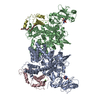
Mass: 347.221 Da / Num. of mol.: 2 / Source method: obtained synthetically / Formula: C10H14N5O7P / Feature type: SUBJECT OF INVESTIGATION / Comment: AMP*YM
#4: Chemical
ChemComp-CL / CHLORIDE ION


Mass: 35.453 Da / Num. of mol.: 4 / Source method: obtained synthetically / Formula: Cl
#5: Chemical ChemComp-PG4 / TETRAETHYLENE GLYCOL


Mass: 194.226 Da / Num. of mol.: 2 / Source method: obtained synthetically / Formula: C8H18O5 / Comment: precipitant*YM
#6: Chemical ChemComp-CA / CALCIUM ION


Mass: 40.078 Da / Num. of mol.: 2 / Source method: obtained synthetically / Formula: Ca / Feature type: SUBJECT OF INVESTIGATION

-
Details

Has ligand of interestY

-
Experimental details

-
Experiment

ExperimentMethod: X-RAY DIFFRACTION / Number of used crystals: 1

-
Sample preparation

CrystalDensity Matthews: 2.73 Å3/Da / Density % sol: 60.03 %
Crystal growTemperature: 299 K / Method: vapor diffusion, sitting drop / pH: 7.5 / Details: 25% PEG 3350, 0.2M NaI, 0.1M HEPES 7.5 / PH range: 6.5-9.0

-
Data collection

DiffractionMean temperature: 100 K / Serial crystal experiment: N
Diffraction sourceSource: SYNCHROTRON / Site: SSRF / Beamline: BL17U1 / Wavelength: 0.97918 Å
DetectorType: ADSC QUANTUM 315r / Detector: CCD / Date: Mar 22, 2019
RadiationProtocol: SINGLE WAVELENGTH / Monochromatic (M) / Laue (L): M / Scattering type: x-ray
Radiation wavelengthWavelength: 0.97918 Å / Relative weight: 1
ReflectionResolution: 3.109→132.187 Å / Num. obs: 41924 / % possible obs: 99.8 % / Redundancy: 6.5 % / CC1/2: 0.997 / Net I/σ(I): 2
Reflection shellResolution: 3.11→3.28 Å / Num. unique obs: 2246 / CC1/2: 0.81

-
Processing

Software
NameVersionClassification
PHENIX(1.11.1_2575: ???)refinement
HKL-2000data reduction
HKL-2000data scaling
MOLREPphasing
RefinementMethod to determine structure: SAD / Resolution: 3.109→66.094 Å / SU ML: 0.4 / Cross valid method: FREE R-VALUE / σ(F): 1.36 / Phase error: 31.06 / Stereochemistry target values: ML
RfactorNum. reflection% reflection
Rfree0.2789 1981 4.73 %
Rwork0.2391 --
obs0.2411 41876 92.46 %
Solvent computationShrinkage radii: 0.9 Å / VDW probe radii: 1.11 Å / Solvent model: FLAT BULK SOLVENT MODEL
Refinement stepCycle: LAST / Resolution: 3.109→66.094 Å
ProteinNucleic acidLigandSolventTotal
Num. atoms12710 0 6 0 12716
Refine LS restraints
Refine-IDTypeDev idealNumber
X-RAY DIFFRACTIONf_bond_d0.00212978
X-RAY DIFFRACTIONf_angle_d0.56517648
X-RAY DIFFRACTIONf_dihedral_angle_d4.9677744
X-RAY DIFFRACTIONf_chiral_restr0.0372038
X-RAY DIFFRACTIONf_plane_restr0.0032266
LS refinement shell
Resolution (Å)Rfactor RfreeNum. reflection RfreeRfactor RworkNum. reflection RworkRefine-ID% reflection obs (%)
3.1085-3.18630.36061450.33713089X-RAY DIFFRACTION100
3.1863-3.27240.36961240.34723072X-RAY DIFFRACTION100
3.2724-3.36870.44581590.33693066X-RAY DIFFRACTION100
3.3687-3.47740.3547340.32391166X-RAY DIFFRACTION37
3.4774-3.60170.32461680.27643074X-RAY DIFFRACTION100
3.6017-3.74590.32671280.25922307X-RAY DIFFRACTION76
3.7459-3.91640.29921410.24472507X-RAY DIFFRACTION82
3.9164-4.12280.27131590.23333072X-RAY DIFFRACTION100
4.1228-4.38110.23841450.21193112X-RAY DIFFRACTION100
4.3811-4.71930.27151570.2093063X-RAY DIFFRACTION100
4.7193-5.1940.27471270.21223087X-RAY DIFFRACTION100
5.194-5.94520.27721780.23613076X-RAY DIFFRACTION100
5.9452-7.48860.23711340.24043133X-RAY DIFFRACTION100
7.4886-66.10850.22951820.2023071X-RAY DIFFRACTION99
Refinement TLS params.Method: refined / Origin x: -3.6447 Å / Origin y: -32.6172 Å / Origin z: -21.2017 Å
111213212223313233
T0.316 Å2-0.0055 Å2-0.0582 Å2-0.365 Å20.0082 Å2--0.4563 Å2
L0.3435 °2-0.022 °2-0.3522 °2-0.2885 °2-0.0312 °2--0.9985 °2
S-0.0114 Å °-0.0261 Å °-0.123 Å °-0.0182 Å °-0.0373 Å °-0.0147 Å °-0.0749 Å °-0.0078 Å °0.0531 Å °
Refinement TLS groupSelection details: all

+
About Yorodumi

-
News

-
Feb 9, 2022. New format data for meta-information of EMDB entries

New format data for meta-information of EMDB entries

  • Version 3 of the EMDB header file is now the official format.
  • The previous official version 1.9 will be removed from the archive.

Related info.:EMDB header

External links:wwPDB to switch to version 3 of the EMDB data model

-
Aug 12, 2020. Covid-19 info

Covid-19 info

URL: https://pdbj.org/emnavi/covid19.php

New page: Covid-19 featured information page in EM Navigator.

Related info.:Covid-19 info / Mar 5, 2020. Novel coronavirus structure data

+
Mar 5, 2020. Novel coronavirus structure data

Novel coronavirus structure data

Related info.:Yorodumi Speices / Aug 12, 2020. Covid-19 info

External links:COVID-19 featured content - PDBj / Molecule of the Month (242):Coronavirus Proteases

+
Jan 31, 2019. EMDB accession codes are about to change! (news from PDBe EMDB page)

EMDB accession codes are about to change! (news from PDBe EMDB page)

  • The allocation of 4 digits for EMDB accession codes will soon come to an end. Whilst these codes will remain in use, new EMDB accession codes will include an additional digit and will expand incrementally as the available range of codes is exhausted. The current 4-digit format prefixed with “EMD-” (i.e. EMD-XXXX) will advance to a 5-digit format (i.e. EMD-XXXXX), and so on. It is currently estimated that the 4-digit codes will be depleted around Spring 2019, at which point the 5-digit format will come into force.
  • The EM Navigator/Yorodumi systems omit the EMD- prefix.

Related info.:Q: What is EMD? / ID/Accession-code notation in Yorodumi/EM Navigator

External links:EMDB Accession Codes are Changing Soon! / Contact to PDBj

+
Jul 12, 2017. Major update of PDB

Major update of PDB

  • wwPDB released updated PDB data conforming to the new PDBx/mmCIF dictionary.
  • This is a major update changing the version number from 4 to 5, and with Remediation, in which all the entries are updated.
  • In this update, many items about electron microscopy experimental information are reorganized (e.g. em_software).
  • Now, EM Navigator and Yorodumi are based on the updated data.

External links:wwPDB Remediation / Enriched Model Files Conforming to OneDep Data Standards Now Available in the PDB FTP Archive

-
Yorodumi

Thousand views of thousand structures

  • Yorodumi is a browser for structure data from EMDB, PDB, SASBDB, etc.
  • This page is also the successor to EM Navigator detail page, and also detail information page/front-end page for Omokage search.
  • The word "yorodu" (or yorozu) is an old Japanese word meaning "ten thousand". "mi" (miru) is to see.

Related info.:EMDB / PDB / SASBDB / Comparison of 3 databanks / Yorodumi Search / Aug 31, 2016. New EM Navigator & Yorodumi / Yorodumi Papers / Jmol/JSmol / Function and homology information / Changes in new EM Navigator and Yorodumi

Read more