 Movie
Movie Controller


+ Open data

- Basic information
| Entry | Database: PDB / ID: 6k32 | |||||||||||||||||||||||||||
|---|---|---|---|---|---|---|---|---|---|---|---|---|---|---|---|---|---|---|---|---|---|---|---|---|---|---|---|---|
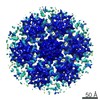
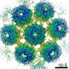
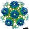
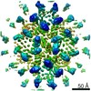
| Title | RdRp complex | |||||||||||||||||||||||||||
 Components Components |
| |||||||||||||||||||||||||||
 Keywords Keywords | VIRAL PROTEIN/RNA / Cypovirus / Transcription / RNA-dependent RNA polymerase / VIRAL PROTEIN-RNA complex | |||||||||||||||||||||||||||
| Function / homology |  Function and homology information Function and homology informationT=2 icosahedral viral capsid / viral inner capsid / viral genome replication / RNA-directed RNA polymerase / RNA-directed RNA polymerase activity / RNA binding Similarity search - Function | |||||||||||||||||||||||||||
| Biological species |  Cypovirus 1 Cypovirus 1 | |||||||||||||||||||||||||||
| Method | ELECTRON MICROSCOPY / single particle reconstruction / cryo EM / Resolution: 3.2 Å | |||||||||||||||||||||||||||
 Authors Authors | Li, X.W. | |||||||||||||||||||||||||||
 Citation Citation | Journal: J Mol Biol / Year: 2020 Title: Structure of RdRps Within a Transcribing dsRNA Virus Provides Insights Into the Mechanisms of RNA Synthesis. Authors: Xiaowu Li / Li Wang / Xurong Wang / Wenyuan Chen / Tao Yang / Jingdong Song / Hongrong Liu / Lingpeng Cheng /  Abstract: RNA-dependent RNA polymerases (RdRps) catalyze RNA synthesis of RNA viruses. During initiation of RNA synthesis, the RdRp catalyzes the formation of the first dinucleotide, acting as primer for ...RNA-dependent RNA polymerases (RdRps) catalyze RNA synthesis of RNA viruses. During initiation of RNA synthesis, the RdRp catalyzes the formation of the first dinucleotide, acting as primer for subsequent processive RNA elongation. Here, we present the structure of the RdRp complexes in the dinucleotide primed state in situ within a transcribing cypovirus under near physiological conditions using cryo-electron microscopy. The 3' end of RNA templates, paired RNA dinucleotide primer, incoming nucleotide, and catalytic divalent cations in the RdRp were resolved at 3.8 Å resolution. The end of the RNA template and the dinucleotide is buttressed by the aromatic tyrosine in a loop from the RdRp bracelet domain. Our structure reveals the interactions between the nucleotide substrates and the conserved residues during the RdRp initiation, and the coordinated structural changes preceding the elongation stage. In addition, it provides the direct evidence for existence of the slow step of the dinucleotide primed state in the viral RdRp transcription. | |||||||||||||||||||||||||||
| History |
|
- Structure visualization
| Movie |
Movie viewer |
|---|---|
| Structure viewer | Molecule: MolmilJmol/JSmol |
- Downloads & links
-Download
| PDBx/mmCIF format | 6k32.cif.gz | 1.3 MB | Display | PDBx/mmCIF format |
|---|---|---|---|---|
| PDB format | pdb6k32.ent.gz | 1.1 MB | Display | PDB format |
| PDBx/mmJSON format | 6k32.json.gz | Tree view | PDBx/mmJSON format | |
| Others |  Other downloads Other downloads |
-Validation report
| Arichive directory | https://data.pdbj.org/pub/pdb/validation_reports/k3/6k32ftp://data.pdbj.org/pub/pdb/validation_reports/k3/6k32 | HTTPS FTP |
|---|
-Related structure data
| Related structure data |  9907MC M: map data used to model this data C: citing same article ( |
|---|---|
| Similar structure data |

-Links
 PDBj
PDBj
- Assembly
| Deposited unit | 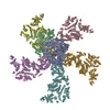
|
|---|---|
| 1 |
|
-Components
-Protein , 5 types, 7 molecules ABCDEFG
| #1: Protein | Mass: 137058.922 Da / Num. of mol.: 1 Source method: isolated from a genetically manipulated source Source: (gene. exp.) Cypovirus 1Production host:  invertebrate environmental sample (environmental samples) invertebrate environmental sample (environmental samples)References: UniProt: D0EZK6 | ||
|---|---|---|---|
| #2: Protein | Mass: 63379.449 Da / Num. of mol.: 1 Source method: isolated from a genetically manipulated source Source: (gene. exp.) Cypovirus 1 / Gene: VP4Production host: invertebrate environmental sample (environmental samples)References: UniProt: C7EWL9 | ||
| #5: Protein | Mass: 136769.422 Da / Num. of mol.: 1 Source method: isolated from a genetically manipulated source Source: (gene. exp.) Cypovirus 1Production host: invertebrate environmental sample (environmental samples)References: UniProt: D3JWE6, UniProt: Q6TS43*PLUS | ||
| #6: Protein | Mass: 135002.609 Da / Num. of mol.: 3 Source method: isolated from a genetically manipulated source Source: (gene. exp.) Cypovirus 1Production host: invertebrate environmental sample (environmental samples)References: UniProt: D3JWE6, UniProt: Q6TS43*PLUS #7: Protein | | Mass: 137422.234 Da / Num. of mol.: 1 Source method: isolated from a genetically manipulated source Source: (gene. exp.) Cypovirus 1Production host: invertebrate environmental sample (environmental samples)References: UniProt: D3JWE6, UniProt: Q6TS43*PLUS |
-RNA chain , 2 types, 2 molecules tp
| #3: RNA chain | Mass: 1507.928 Da / Num. of mol.: 1 Source method: isolated from a genetically manipulated source Source: (gene. exp.) Cypovirus 1Production host: invertebrate environmental sample (environmental samples) |
|---|---|
| #4: RNA chain | Mass: 869.394 Da / Num. of mol.: 1 Source method: isolated from a genetically manipulated source Source: (gene. exp.) Cypovirus 1Production host: invertebrate environmental sample (environmental samples) |
-Non-polymers , 5 types, 6 molecules 


| #8: Chemical |  Mass: 24.305 Da / Num. of mol.: 2 / Source method: obtained synthetically / Formula: Mg Mass: 24.305 Da / Num. of mol.: 2 / Source method: obtained synthetically / Formula: Mg#9: Chemical | ChemComp-MG7 / |  Mass: 298.275 Da / Num. of mol.: 1 / Source method: obtained synthetically / Formula: C11H16N5O5 Mass: 298.275 Da / Num. of mol.: 1 / Source method: obtained synthetically / Formula: C11H16N5O5#10: Chemical | ChemComp-DPO / |  Mass: 173.943 Da / Num. of mol.: 1 / Source method: obtained synthetically / Formula: O7P2 Mass: 173.943 Da / Num. of mol.: 1 / Source method: obtained synthetically / Formula: O7P2#11: Chemical | ChemComp-A2M / |  Type: RNA linking / Mass: 361.248 Da / Num. of mol.: 1 / Source method: obtained synthetically / Formula: C11H16N5O7P Type: RNA linking / Mass: 361.248 Da / Num. of mol.: 1 / Source method: obtained synthetically / Formula: C11H16N5O7P#12: Chemical | ChemComp-UTP / |  Mass: 484.141 Da / Num. of mol.: 1 / Source method: obtained synthetically / Formula: C9H15N2O15P3 / Comment: UTP*YM Mass: 484.141 Da / Num. of mol.: 1 / Source method: obtained synthetically / Formula: C9H15N2O15P3 / Comment: UTP*YM |
|---|
-Details
| Has ligand of interest | N |
|---|---|
| Has protein modification | N |
-Experimental details
-Experiment
| Experiment | Method: ELECTRON MICROSCOPY |
|---|---|
| EM experiment | Aggregation state: PARTICLE / 3D reconstruction method: single particle reconstruction |
- Sample preparation
| Component | Name: Cypovirus / Type: VIRUS / Entity ID: #1-#7 / Source: NATURAL |
|---|---|
| Source (natural) | Organism: Cypovirus (cytoplasmic polyhedrosis viruses) |
| Details of virus | Empty: NO / Enveloped: NO / Isolate: OTHER / Type: VIRION |
| Buffer solution | pH: 8 |
| Specimen | Embedding applied: NO / Shadowing applied: NO / Staining applied: NO / Vitrification applied: YES |
| Vitrification | Cryogen name: OTHER |
- Electron microscopy imaging
| Experimental equipment |  Model: Talos Arctica / Image courtesy: FEI Company |
|---|---|
| Microscopy | Model: FEI TECNAI ARCTICA |
| Electron gun | Electron source:  FIELD EMISSION GUN / Accelerating voltage: 200 kV / Illumination mode: FLOOD BEAM FIELD EMISSION GUN / Accelerating voltage: 200 kV / Illumination mode: FLOOD BEAM |
| Electron lens | Mode: BRIGHT FIELD |
| Image recording | Electron dose: 20 e/Å2 / Film or detector model: FEI FALCON II (4k x 4k) |
- Processing
| Software | Name: PHENIX / Version: 1.13_2998: / Classification: refinement |
|---|---|
| EM software | Name: PHENIX / Category: model refinement |
| CTF correction | Type: PHASE FLIPPING AND AMPLITUDE CORRECTION |
| 3D reconstruction | Resolution: 3.2 Å / Resolution method: FSC 0.143 CUT-OFF / Num. of particles: 100000 / Symmetry type: POINT |
| Refinement | Highest resolution: 3.2 Å |
