 Movie
Movie Controller


+ Open data

- Basic information
| Entry | Database: PDB / ID: 6jyx | |||||||||
|---|---|---|---|---|---|---|---|---|---|---|
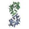
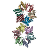
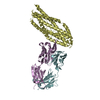
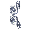
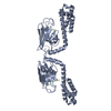
| Title | Structure of CbpJ from Streptococcus Pneumoniae TIGR4 | |||||||||
 Components Components | Choline binding protein J | |||||||||
 Keywords Keywords | CHOLINE-BINDING PROTEIN / Streptococcus pneumoniae / choline-binding proteins / adhesion | |||||||||
| Function / homology |  Function and homology information Function and homology informationleft handed beta-beta-3-solenoid - #20 / left handed beta-beta-3-solenoid / Choline-binding repeat / Putative cell wall binding repeat / Cell wall/choline-binding repeat / Cell wall-binding repeat profile. / Ribbon / Mainly Beta Similarity search - Domain/homology | |||||||||
| Biological species |  Streptococcus pneumoniae serotype 4 (bacteria) Streptococcus pneumoniae serotype 4 (bacteria) | |||||||||
| Method |  X-RAY DIFFRACTION / SYNCHROTRON / MOLECULAR REPLACEMENT / Resolution: 2 Å X-RAY DIFFRACTION / SYNCHROTRON / MOLECULAR REPLACEMENT / Resolution: 2 Å | |||||||||
 Authors Authors | Xu, Q. / Zhang, J.W. / Li, Q. / Jiang, Y.L. | |||||||||
| Funding support |  China, 2items China, 2items
| |||||||||
 Citation Citation | Journal: Biochem.Biophys.Res.Commun. / Year: 2019 Title: Crystal structure of the choline-binding protein CbpJ from Streptococcus pneumoniae. Authors: Xu, Q. / Zhang, J.W. / Chen, Y. / Li, Q. / Jiang, Y.L. | |||||||||
| History |
|
- Structure visualization
| Structure viewer | Molecule: MolmilJmol/JSmol |
|---|
- Downloads & links
-Download
| PDBx/mmCIF format | 6jyx.cif.gz | 144.4 KB | Display | PDBx/mmCIF format |
|---|---|---|---|---|
| PDB format | pdb6jyx.ent.gz | 111.8 KB | Display | PDB format |
| PDBx/mmJSON format | 6jyx.json.gz | Tree view | PDBx/mmJSON format | |
| Others |  Other downloads Other downloads |
-Validation report
| Arichive directory | https://data.pdbj.org/pub/pdb/validation_reports/jy/6jyxftp://data.pdbj.org/pub/pdb/validation_reports/jy/6jyx | HTTPS FTP |
|---|
-Related structure data
| Related structure data | 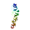 2v04S S: Starting model for refinement |
|---|---|
| Similar structure data |


-Links
 PDBj
PDBj
- Assembly
| Deposited unit | 
| ||||||||
|---|---|---|---|---|---|---|---|---|---|
| 1 |
| ||||||||
| Unit cell |
|
-Components
| #1: Protein | Mass: 35787.031 Da / Num. of mol.: 2 Source method: isolated from a genetically manipulated source Source: (gene. exp.) Streptococcus pneumoniae serotype 4 (strain ATCC BAA-334 / TIGR4) (bacteria)Strain: ATCC BAA-334 / TIGR4 / Gene: cbpJ, SP_0378 / Plasmid: pET-28a-derived / Production host: #2: Chemical | ChemComp-CHT /   Mass: 104.171 Da / Num. of mol.: 14 / Source method: obtained synthetically / Formula: C5H14NO Mass: 104.171 Da / Num. of mol.: 14 / Source method: obtained synthetically / Formula: C5H14NO#3: Chemical |   Mass: 106.120 Da / Num. of mol.: 2 / Source method: obtained synthetically / Formula: C4H10O3 Mass: 106.120 Da / Num. of mol.: 2 / Source method: obtained synthetically / Formula: C4H10O3#4: Water | ChemComp-HOH / |  Mass: 18.015 Da / Num. of mol.: 319 / Source method: isolated from a natural source / Formula: H2O Mass: 18.015 Da / Num. of mol.: 319 / Source method: isolated from a natural source / Formula: H2O |
|---|
-Experimental details
-Experiment
| Experiment | Method: X-RAY DIFFRACTION / Number of used crystals: 1 |
|---|
- Sample preparation
| Crystal | Density Matthews: 2.77 Å3/Da / Density % sol: 55.57 % |
|---|---|
| Crystal grow | Temperature: 289 K / Method: vapor diffusion, hanging drop / pH: 6.5 / Details: 10% PEG 20K, 0.1 M MES pH 6.5 |
-Data collection
| Diffraction | Mean temperature: 100 K / Serial crystal experiment: N |
|---|---|
| Diffraction source | Source: SYNCHROTRON / Site: SSRF / Beamline: BL17U / Wavelength: 0.97923 Å |
| Detector | Type: MARRESEARCH / Detector: CCD / Date: Jun 7, 2010 |
| Radiation | Protocol: SINGLE WAVELENGTH / Monochromatic (M) / Laue (L): M / Scattering type: x-ray |
| Radiation wavelength | Wavelength: 0.97923 Å / Relative weight: 1 |
| Reflection | Resolution: 2→50 Å / Num. obs: 51107 / % possible obs: 93.4 % / Redundancy: 6.6 % / Rmerge(I) obs: 0.072 / Net I/σ(I): 10.259 |
| Reflection shell | Resolution: 2→2.07 Å / Rmerge(I) obs: 0.199 / Num. unique obs: 1000 |
- Processing
| Software |
| ||||||||||||||||||||||||||||||||||||||||||||||||||||||||||||||||||||||||||||||||||||||||||||||||||||||||||||||||||||||||||||||||||||||||||||||||||||||||||||||||||||||||||||||||||||||
|---|---|---|---|---|---|---|---|---|---|---|---|---|---|---|---|---|---|---|---|---|---|---|---|---|---|---|---|---|---|---|---|---|---|---|---|---|---|---|---|---|---|---|---|---|---|---|---|---|---|---|---|---|---|---|---|---|---|---|---|---|---|---|---|---|---|---|---|---|---|---|---|---|---|---|---|---|---|---|---|---|---|---|---|---|---|---|---|---|---|---|---|---|---|---|---|---|---|---|---|---|---|---|---|---|---|---|---|---|---|---|---|---|---|---|---|---|---|---|---|---|---|---|---|---|---|---|---|---|---|---|---|---|---|---|---|---|---|---|---|---|---|---|---|---|---|---|---|---|---|---|---|---|---|---|---|---|---|---|---|---|---|---|---|---|---|---|---|---|---|---|---|---|---|---|---|---|---|---|---|---|---|---|---|
| Refinement | Method to determine structure: MOLECULAR REPLACEMENT Starting model: 2V04 Resolution: 2→48.68 Å / Cor.coef. Fo:Fc: 0.953 / Cor.coef. Fo:Fc free: 0.933 / SU B: 4.108 / SU ML: 0.117 / Cross valid method: THROUGHOUT / ESU R: 0.198 / ESU R Free: 0.177 / Details: HYDROGENS HAVE BEEN ADDED IN THE RIDING POSITIONS
| ||||||||||||||||||||||||||||||||||||||||||||||||||||||||||||||||||||||||||||||||||||||||||||||||||||||||||||||||||||||||||||||||||||||||||||||||||||||||||||||||||||||||||||||||||||||
| Solvent computation | Ion probe radii: 0.8 Å / Shrinkage radii: 0.8 Å / VDW probe radii: 1.4 Å | ||||||||||||||||||||||||||||||||||||||||||||||||||||||||||||||||||||||||||||||||||||||||||||||||||||||||||||||||||||||||||||||||||||||||||||||||||||||||||||||||||||||||||||||||||||||
| Displacement parameters | Biso mean: 42.374 Å2
| ||||||||||||||||||||||||||||||||||||||||||||||||||||||||||||||||||||||||||||||||||||||||||||||||||||||||||||||||||||||||||||||||||||||||||||||||||||||||||||||||||||||||||||||||||||||
| Refinement step | Cycle: 1 / Resolution: 2→48.68 Å
| ||||||||||||||||||||||||||||||||||||||||||||||||||||||||||||||||||||||||||||||||||||||||||||||||||||||||||||||||||||||||||||||||||||||||||||||||||||||||||||||||||||||||||||||||||||||
| Refine LS restraints |
|