 Movie
Movie Controller

[English] 日本語
 Yorodumi
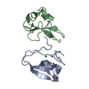
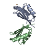
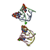
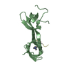
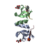
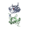
- PDB-6jui: The atypical Myb-like protein Cdc5 contains two distinct nucleic ... -
+ Open data

- Basic information
| Entry | Database: PDB / ID: 6jui | ||||||
|---|---|---|---|---|---|---|---|
| Title | The atypical Myb-like protein Cdc5 contains two distinct nucleic acid-binding surfaces | ||||||
 Components Components | Pre-mRNA-splicing factor CEF1 | ||||||
 Keywords Keywords | DNA BINDING PROTEIN / crystal / DNA binding domain / Myb domain / rice blast fungus / RNA binding | ||||||
| Function / homology |  Function and homology information Function and homology informationnucleolar peripheral inclusion body / post-mRNA release spliceosomal complex / mRNA cis splicing, via spliceosome / Prp19 complex / DNA binding / cytosol Similarity search - Function | ||||||
| Biological species |  Magnaporthe oryzae (rice blast fungus) Magnaporthe oryzae (rice blast fungus) | ||||||
| Method |  X-RAY DIFFRACTION / SYNCHROTRON / MOLECULAR REPLACEMENT / Resolution: 2.402 Å X-RAY DIFFRACTION / SYNCHROTRON / MOLECULAR REPLACEMENT / Resolution: 2.402 Å | ||||||
 Authors Authors | Wang, C. / Li, G. / Li, M. / Yang, J. / Liu, J. | ||||||
 Citation Citation | Journal: Biochem.J. / Year: 2019 Title: Two distinct nucleic acid binding surfaces of Cdc5 regulate development. Authors: Wang, C. / Li, M. / Li, G. / Liu, X. / Zhao, W. / Yu, B. / Liu, J. / Yang, J. / Peng, Y.L. | ||||||
| History |
|
- Structure visualization
| Structure viewer | Molecule: MolmilJmol/JSmol |
|---|
- Downloads & links
-Download
| PDBx/mmCIF format | 6jui.cif.gz | 56.4 KB | Display | PDBx/mmCIF format |
|---|---|---|---|---|
| PDB format | pdb6jui.ent.gz | 40.7 KB | Display | PDB format |
| PDBx/mmJSON format | 6jui.json.gz | Tree view | PDBx/mmJSON format | |
| Others |  Other downloads Other downloads |
-Validation report
| Summary document | 6jui_validation.pdf.gz | 425.2 KB | Display | wwPDB validaton report |
|---|---|---|---|---|
| Full document | 6jui_full_validation.pdf.gz | 427.7 KB | Display | |
| Data in XML | 6jui_validation.xml.gz | 6.2 KB | Display | |
| Data in CIF | 6jui_validation.cif.gz | 7.2 KB | Display | |
| Arichive directory | https://data.pdbj.org/pub/pdb/validation_reports/ju/6juiftp://data.pdbj.org/pub/pdb/validation_reports/ju/6jui | HTTPS FTP |
-Related structure data
| Similar structure data |
|---|


-Links
 PDBj
PDBj
- Assembly
| Deposited unit | 
| ||||||||
|---|---|---|---|---|---|---|---|---|---|
| 1 |
| ||||||||
| Unit cell |
|
-Components
| #1: Protein | Mass: 13507.555 Da / Num. of mol.: 1 Source method: isolated from a genetically manipulated source Source: (gene. exp.) Magnaporthe oryzae (rice blast fungus) / Gene: CEF1, MGG_01426 / Production host:  |
|---|---|
| #2: Water | ChemComp-HOH /  Mass: 18.015 Da / Num. of mol.: 2 / Source method: isolated from a natural source / Formula: H2O Mass: 18.015 Da / Num. of mol.: 2 / Source method: isolated from a natural source / Formula: H2O |
-Experimental details
-Experiment
| Experiment | Method: X-RAY DIFFRACTION / Number of used crystals: 1 |
|---|
- Sample preparation
| Crystal | Density Matthews: 2.8 Å3/Da / Density % sol: 56 % |
|---|---|
| Crystal grow | Temperature: 291 K / Method: vapor diffusion, sitting drop / Details: 40% MPD |
-Data collection
| Diffraction | Mean temperature: 100 K / Serial crystal experiment: N |
|---|---|
| Diffraction source | Source: SYNCHROTRON / Site: SSRF  / Beamline: BL17U / Wavelength: 0.9792 Å / Beamline: BL17U / Wavelength: 0.9792 Å |
| Detector | Type: ADSC QUANTUM 315r / Detector: CCD / Date: Apr 12, 2016 |
| Radiation | Protocol: SINGLE WAVELENGTH / Monochromatic (M) / Laue (L): M / Scattering type: x-ray |
| Radiation wavelength | Wavelength: 0.9792 Å / Relative weight: 1 |
| Reflection | Resolution: 2.402→38.14 Å / Num. obs: 11354 / % possible obs: 99.29 % / Redundancy: 7.4 % / Net I/σ(I): 22.1 |
| Reflection shell | Resolution: 2.402→2.488 Å |
- Processing
| Software |
| |||||||||||||||||||||||||||||||||||||||||||||||||||||||||||||||||||||||||||||||||||||||||||||||||||||||||||||||||||||||||||||||||||||||||||||||||||||||||||||||||||||||||||||||
|---|---|---|---|---|---|---|---|---|---|---|---|---|---|---|---|---|---|---|---|---|---|---|---|---|---|---|---|---|---|---|---|---|---|---|---|---|---|---|---|---|---|---|---|---|---|---|---|---|---|---|---|---|---|---|---|---|---|---|---|---|---|---|---|---|---|---|---|---|---|---|---|---|---|---|---|---|---|---|---|---|---|---|---|---|---|---|---|---|---|---|---|---|---|---|---|---|---|---|---|---|---|---|---|---|---|---|---|---|---|---|---|---|---|---|---|---|---|---|---|---|---|---|---|---|---|---|---|---|---|---|---|---|---|---|---|---|---|---|---|---|---|---|---|---|---|---|---|---|---|---|---|---|---|---|---|---|---|---|---|---|---|---|---|---|---|---|---|---|---|---|---|---|---|---|---|---|
| Refinement | Method to determine structure: MOLECULAR REPLACEMENT / Resolution: 2.402→38.139 Å / SU ML: 0.25 / Cross valid method: FREE R-VALUE / σ(F): 1.31 / Phase error: 34.12
| |||||||||||||||||||||||||||||||||||||||||||||||||||||||||||||||||||||||||||||||||||||||||||||||||||||||||||||||||||||||||||||||||||||||||||||||||||||||||||||||||||||||||||||||
| Solvent computation | Shrinkage radii: 0.9 Å / VDW probe radii: 1.11 Å | |||||||||||||||||||||||||||||||||||||||||||||||||||||||||||||||||||||||||||||||||||||||||||||||||||||||||||||||||||||||||||||||||||||||||||||||||||||||||||||||||||||||||||||||
| Refinement step | Cycle: LAST / Resolution: 2.402→38.139 Å
| |||||||||||||||||||||||||||||||||||||||||||||||||||||||||||||||||||||||||||||||||||||||||||||||||||||||||||||||||||||||||||||||||||||||||||||||||||||||||||||||||||||||||||||||
| Refine LS restraints |
| |||||||||||||||||||||||||||||||||||||||||||||||||||||||||||||||||||||||||||||||||||||||||||||||||||||||||||||||||||||||||||||||||||||||||||||||||||||||||||||||||||||||||||||||
| LS refinement shell |
| |||||||||||||||||||||||||||||||||||||||||||||||||||||||||||||||||||||||||||||||||||||||||||||||||||||||||||||||||||||||||||||||||||||||||||||||||||||||||||||||||||||||||||||||
| Refinement TLS params. | Method: refined / Refine-ID: X-RAY DIFFRACTION
| |||||||||||||||||||||||||||||||||||||||||||||||||||||||||||||||||||||||||||||||||||||||||||||||||||||||||||||||||||||||||||||||||||||||||||||||||||||||||||||||||||||||||||||||
| Refinement TLS group |
|
