 Movie
Movie Controller

[English] 日本語
 Yorodumi
- PDB-6ju9: Aspergillus oryzae active-tyrosinase copper-bound C92A mutant com... -
+ Open data

- Basic information
| Entry | Database: PDB / ID: 6ju9 | |||||||||
|---|---|---|---|---|---|---|---|---|---|---|
| Title | Aspergillus oryzae active-tyrosinase copper-bound C92A mutant complexed with L-tyrosine | |||||||||
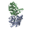
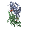
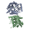
 Components Components | Tyrosinase | |||||||||
 Keywords Keywords | OXIDOREDUCTASE / Tyrosinase / Copper enzyme / dinuclear copper center | |||||||||
| Function / homology |  Function and homology information Function and homology informationtyrosinase / tyrosinase activity / melanin biosynthetic process / metal ion binding / nucleus Similarity search - Function | |||||||||
| Biological species |  | |||||||||
| Method |  X-RAY DIFFRACTION / SYNCHROTRON / MOLECULAR REPLACEMENT / Resolution: 1.42 Å X-RAY DIFFRACTION / SYNCHROTRON / MOLECULAR REPLACEMENT / Resolution: 1.42 Å | |||||||||
 Authors Authors | Fujieda, N. / Umakoshi, K. / Nishikawa, Y. / Kurisu, G. / Itoh, S. | |||||||||
| Funding support |  Japan, 1items Japan, 1items
| |||||||||
 Citation Citation | Journal: Angew.Chem.Int.Ed.Engl. / Year: 2020 Title: Copper-Oxygen Dynamics in the Tyrosinase Mechanism. Authors: Fujieda, N. / Umakoshi, K. / Ochi, Y. / Nishikawa, Y. / Yanagisawa, S. / Kubo, M. / Kurisu, G. / Itoh, S. | |||||||||
| History |
|
- Structure visualization
| Structure viewer | Molecule: MolmilJmol/JSmol |
|---|
- Downloads & links
-Download
| PDBx/mmCIF format | 6ju9.cif.gz | 375.9 KB | Display | PDBx/mmCIF format |
|---|---|---|---|---|
| PDB format | pdb6ju9.ent.gz | 301.1 KB | Display | PDB format |
| PDBx/mmJSON format | 6ju9.json.gz | Tree view | PDBx/mmJSON format | |
| Others |  Other downloads Other downloads |
-Validation report
| Arichive directory | https://data.pdbj.org/pub/pdb/validation_reports/ju/6ju9ftp://data.pdbj.org/pub/pdb/validation_reports/ju/6ju9 | HTTPS FTP |
|---|
-Related structure data
| Related structure data |  6ju4C  6ju5C 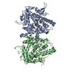 6ju6C  6ju7C  6ju8C 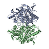 6juaC 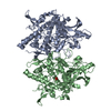 6jubC 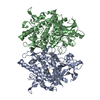 6jucC 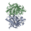 6judC 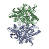 3w6wS S: Starting model for refinement C: citing same article ( |
|---|---|
| Similar structure data |


-Links
 PDBj
PDBj

- Assembly
| Deposited unit | 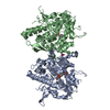
| ||||||||
|---|---|---|---|---|---|---|---|---|---|
| 1 |
| ||||||||
| Unit cell |
|
-Components
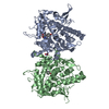
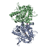
-Protein , 1 types, 2 molecules AB
| #1: Protein | Mass: 53495.855 Da / Num. of mol.: 2 / Mutation: C92A Source method: isolated from a genetically manipulated source Source: (gene. exp.)  |
|---|
-Non-polymers , 5 types, 751 molecules 
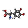


| #2: Chemical |  Type: L-peptide linking / Mass: 181.189 Da / Num. of mol.: 2 / Source method: obtained synthetically / Formula: C9H11NO3 Type: L-peptide linking / Mass: 181.189 Da / Num. of mol.: 2 / Source method: obtained synthetically / Formula: C9H11NO3#3: Chemical | ChemComp-DAH / |  Type: L-peptide linking / Mass: 197.188 Da / Num. of mol.: 1 / Source method: obtained synthetically / Formula: C9H11NO4 Type: L-peptide linking / Mass: 197.188 Da / Num. of mol.: 1 / Source method: obtained synthetically / Formula: C9H11NO4#4: Chemical | ChemComp-CU /  Mass: 63.546 Da / Num. of mol.: 4 / Source method: obtained synthetically / Formula: Cu Mass: 63.546 Da / Num. of mol.: 4 / Source method: obtained synthetically / Formula: Cu#5: Chemical | ChemComp-NO3 / |  Mass: 62.005 Da / Num. of mol.: 1 / Source method: obtained synthetically / Formula: NO3 Mass: 62.005 Da / Num. of mol.: 1 / Source method: obtained synthetically / Formula: NO3#6: Water | ChemComp-HOH / | Mass: 18.015 Da / Num. of mol.: 743 / Source method: isolated from a natural source / Formula: H2O |
|---|
-Details
| Has protein modification | N |
|---|
-Experimental details
-Experiment
| Experiment | Method: X-RAY DIFFRACTION / Number of used crystals: 1 |
|---|
- Sample preparation
| Crystal | Density Matthews: 2.07 Å3/Da / Density % sol: 40.64 % |
|---|---|
| Crystal grow | Temperature: 293 K / Method: vapor diffusion, hanging drop / pH: 7.2 / Details: 20% polyethylene glycol (PEG) 3350, 30mM NH4NO3 |
-Data collection
| Diffraction | Mean temperature: 100 K / Serial crystal experiment: N |
|---|---|
| Diffraction source | Source: SYNCHROTRON / Site: SPring-8 / Beamline: BL44XU / Wavelength: 0.9 Å |
| Detector | Type: RAYONIX MX300HE / Detector: CCD / Date: Jun 12, 2015 |
| Radiation | Protocol: SINGLE WAVELENGTH / Monochromatic (M) / Laue (L): M / Scattering type: x-ray |
| Radiation wavelength | Wavelength: 0.9 Å / Relative weight: 1 |
| Reflection | Resolution: 1.42→50 Å / Num. obs: 164029 / % possible obs: 97.9 % / Redundancy: 7 % / Rmerge(I) obs: 0.087 / Net I/σ(I): 19.2 |
| Reflection shell | Resolution: 1.42→1.44 Å / Redundancy: 7 % / Rmerge(I) obs: 0.39 / Mean I/σ(I) obs: 5.2 / Num. unique obs: 7964 / % possible all: 96 |
- Processing
| Software |
| |||||||||||||||||||||||||||||||||
|---|---|---|---|---|---|---|---|---|---|---|---|---|---|---|---|---|---|---|---|---|---|---|---|---|---|---|---|---|---|---|---|---|---|---|
| Refinement | Method to determine structure: MOLECULAR REPLACEMENT Starting model: 3W6W Resolution: 1.42→30 Å / Cross valid method: FREE R-VALUE / σ(F): 0
| |||||||||||||||||||||||||||||||||
| Refine analyze | Num. disordered residues: 33 / Occupancy sum hydrogen: 0 / Occupancy sum non hydrogen: 7709.1 | |||||||||||||||||||||||||||||||||
| Refinement step | Cycle: LAST / Resolution: 1.42→30 Å
| |||||||||||||||||||||||||||||||||
| Refine LS restraints |
|
