 Movie
Movie Controller


+ Open data

- Basic information
| Entry | Database: PDB / ID: 6jsx | ||||||
|---|---|---|---|---|---|---|---|
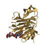
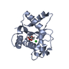
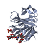
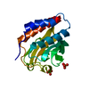
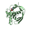
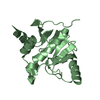
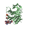
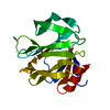
| Title | Structure of a flagellin protein, HpFlaG | ||||||
 Components Components | Flagellar biosynthesis protein FlaG | ||||||
 Keywords Keywords | BIOSYNTHETIC PROTEIN / Helicobacter pylori / flagella | ||||||
| Function / homology | FlaG protein / FlaG-like superfamily / FlaG protein / Flagellar biosynthesis protein FlaG Function and homology information Function and homology information | ||||||
| Biological species |   Helicobacter pylori (bacteria) Helicobacter pylori (bacteria) | ||||||
| Method |  X-RAY DIFFRACTION / SYNCHROTRON / MAD / Resolution: 2.7 Å X-RAY DIFFRACTION / SYNCHROTRON / MAD / Resolution: 2.7 Å | ||||||
 Authors Authors | Tsai, J.Y. / Sun, Y.J. / Hsiao, C.D. | ||||||
| Funding support |  Taiwan, 1items Taiwan, 1items
| ||||||
 Citation Citation | Journal: J Chin Chem Soc / Year: 2019 Title: Crystal structure of the flagellin protein FlaG from Helicobacter pylori. Authors: Tsai, J.Y. / Yeh, Y.H. / Lin, L.D. / Sun, Y.J. / Hsiao, C.D. | ||||||
| History |
|
- Structure visualization
| Structure viewer | Molecule: MolmilJmol/JSmol |
|---|
- Downloads & links
-Download
| PDBx/mmCIF format | 6jsx.cif.gz | 74.2 KB | Display | PDBx/mmCIF format |
|---|---|---|---|---|
| PDB format | pdb6jsx.ent.gz | 55.9 KB | Display | PDB format |
| PDBx/mmJSON format | 6jsx.json.gz | Tree view | PDBx/mmJSON format | |
| Others |  Other downloads Other downloads |
-Validation report
| Summary document | 6jsx_validation.pdf.gz | 423.5 KB | Display | wwPDB validaton report |
|---|---|---|---|---|
| Full document | 6jsx_full_validation.pdf.gz | 425 KB | Display | |
| Data in XML | 6jsx_validation.xml.gz | 7.9 KB | Display | |
| Data in CIF | 6jsx_validation.cif.gz | 9.9 KB | Display | |
| Arichive directory | https://data.pdbj.org/pub/pdb/validation_reports/js/6jsxftp://data.pdbj.org/pub/pdb/validation_reports/js/6jsx | HTTPS FTP |
-Related structure data
| Similar structure data |
|---|


-Links
 PDBj
PDBj- Assembly
| Deposited unit | 
| ||||||||
|---|---|---|---|---|---|---|---|---|---|
| 1 |
| ||||||||
| Unit cell |
|
-Components
| #1: Protein | Mass: 8438.707 Da / Num. of mol.: 2 Source method: isolated from a genetically manipulated source Source: (gene. exp.) Helicobacter pylori (bacteria) / Gene: BB430_02730, HPY1198_01455 / Production host: #2: Water | ChemComp-HOH / |  Mass: 18.015 Da / Num. of mol.: 41 / Source method: isolated from a natural source / Formula: H2O Mass: 18.015 Da / Num. of mol.: 41 / Source method: isolated from a natural source / Formula: H2O |
|---|
-Experimental details
-Experiment
| Experiment | Method: X-RAY DIFFRACTION / Number of used crystals: 1 |
|---|
- Sample preparation
| Crystal | Density Matthews: 3.38 Å3/Da / Density % sol: 63.65 % |
|---|---|
| Crystal grow | Temperature: 293 K / Method: vapor diffusion, hanging drop / Details: 28% (NH4)2SO4, 0.1M K/Na Tartrate |
-Data collection
| Diffraction | Mean temperature: 100 K / Serial crystal experiment: N | |||||||||||||||||||||||||||||||||||||||||||||||||||||||||||||||||||||||||||||||||||||||||||||||||||
|---|---|---|---|---|---|---|---|---|---|---|---|---|---|---|---|---|---|---|---|---|---|---|---|---|---|---|---|---|---|---|---|---|---|---|---|---|---|---|---|---|---|---|---|---|---|---|---|---|---|---|---|---|---|---|---|---|---|---|---|---|---|---|---|---|---|---|---|---|---|---|---|---|---|---|---|---|---|---|---|---|---|---|---|---|---|---|---|---|---|---|---|---|---|---|---|---|---|---|---|---|
| Diffraction source | Source: SYNCHROTRON / Site: NSRRC / Beamline: BL13B1 / Wavelength: 0.9641 Å | |||||||||||||||||||||||||||||||||||||||||||||||||||||||||||||||||||||||||||||||||||||||||||||||||||
| Detector | Type: ADSC QUANTUM 315 / Detector: CCD / Date: Mar 30, 2006 | |||||||||||||||||||||||||||||||||||||||||||||||||||||||||||||||||||||||||||||||||||||||||||||||||||
| Radiation | Protocol: SINGLE WAVELENGTH / Monochromatic (M) / Laue (L): M / Scattering type: x-ray | |||||||||||||||||||||||||||||||||||||||||||||||||||||||||||||||||||||||||||||||||||||||||||||||||||
| Radiation wavelength | Wavelength: 0.9641 Å / Relative weight: 1 | |||||||||||||||||||||||||||||||||||||||||||||||||||||||||||||||||||||||||||||||||||||||||||||||||||
| Reflection | Resolution: 2.7→30 Å / Num. obs: 7055 / % possible obs: 98.6 % / Redundancy: 8.2 % / Biso Wilson estimate: 75.72 Å2 / Rmerge(I) obs: 0.054 / Rpim(I) all: 0.02 / Rrim(I) all: 0.058 / Χ2: 1.343 / Net I/σ(I): 14.8 | |||||||||||||||||||||||||||||||||||||||||||||||||||||||||||||||||||||||||||||||||||||||||||||||||||
| Reflection shell | Diffraction-ID: 1
|
- Processing
| Software |
| ||||||||||||||||||||||||||||||||||||||||||
|---|---|---|---|---|---|---|---|---|---|---|---|---|---|---|---|---|---|---|---|---|---|---|---|---|---|---|---|---|---|---|---|---|---|---|---|---|---|---|---|---|---|---|---|
| Refinement | Method to determine structure: MAD / Resolution: 2.7→29.206 Å / SU ML: 0.4 / Cross valid method: THROUGHOUT / σ(F): 1.34 / Phase error: 33.34
| ||||||||||||||||||||||||||||||||||||||||||
| Solvent computation | Shrinkage radii: 0.9 Å / VDW probe radii: 1.11 Å | ||||||||||||||||||||||||||||||||||||||||||
| Displacement parameters | Biso max: 142.92 Å2 / Biso mean: 72.26 Å2 / Biso min: 40.05 Å2 | ||||||||||||||||||||||||||||||||||||||||||
| Refinement step | Cycle: final / Resolution: 2.7→29.206 Å
| ||||||||||||||||||||||||||||||||||||||||||
| Refine LS restraints |
| ||||||||||||||||||||||||||||||||||||||||||
| LS refinement shell | Refine-ID: X-RAY DIFFRACTION / Rfactor Rfree error: 0 / Total num. of bins used: 5
| ||||||||||||||||||||||||||||||||||||||||||
| Refinement TLS params. | Method: refined / Origin x: -13.6135 Å / Origin y: -16.3829 Å / Origin z: -3.7017 Å
| ||||||||||||||||||||||||||||||||||||||||||
| Refinement TLS group |
|
